How MedGenome’s unique next-generation sequencing solutions are helping precision therapies / personalized medicine

Recent advances in next-generation sequencing technologies have heralded a paradigm shift in the field of precision oncology and personalized/genomic medicine, with a large number of somatic- and germline mutation-profiling programs worldwide. These programs have paved the way for personalized medicine in contrast to a unified approach that clearly fails in select individuals, conferring benefits to only a subset of patients. While these genomic analyses become increasingly accessible and almost commonplace to all research scientists, clinicians and molecular geneticists, they are faced with the challenging task of interpreting and translating the results from these analyses.

By Savita Jayaram Ph.D., Kushal Suryamohan Ph.D., MedGenome Scientific Affairs

Recent advances in next-generation sequencing technologies have heralded a paradigm shift in the field of precision oncology and personalized/genomic medicine, with a large number of somatic- and germline mutation-profiling programs worldwide. These programs have paved the way for personalized medicine in contrast to a unified approach that clearly fails in select individuals, conferring benefits to only a subset of patients. While these genomic analyses become increasingly accessible and almost commonplace to all research scientists, clinicians and molecular geneticists, they are faced with the challenging task of interpreting and translating the results from these analyses.

Owing to the inherent heterogeneity and complexity of solid and liquid tumors and inherited cancers, newer bioinformatics tools are required to interpret and prioritize the variants, including SNPs, In-Dels, copy number variations (CNVs), translocations, gene fusions, and splice variants, covering a broad spectrum of genomic alterations. Recently, MedGenome Labs launched the AI-enabled VarMiner pipeline to detect actionable genetic variants in rare and inherited cancers, powered by internally benchmarked tools and databases, to precisely pinpoint these changes with accuracy and efficiency. Identifying these causal variants is like pulling out a needle in a haystack, but is the need of the hour, to improve prediction rate with higher specificity.

Custom Tumor Panels

Further, to provide an easy identification and maximize the utility of these analyses, MedGenome has developed many tumor panels such as TSO170 and TST500. These panels enable fast and seamless reporting ranging from blood-based markers from circulating tumor cells (from liquid biopsies) to high depth sequencing of tumor mutation burden to provide insights into the potential solutions for the patients, in both research and clinical settings. TruSight Oncology 500 offers wide variety of benefits in analyzing multiple tumor variants across 523 genes in a single assay, enabling comprehensive genomic profiling of tumor samples. The assay is highly effective in identifying all types of relevant DNA and RNA variants in different types of solid tumors encompassing sarcomas, lung, melanoma, ovarian, breast, gastric, and bladder cancers. Also, the assay is highly accurate in measuring immuno-oncology biomarkers such as microsatellite instability (MSI) and tumor mutational burden (TMB). Our TST170 Panel had been validated on circulating tumor DNA (ctDNA) providing an in-depth view into cancer genetics.

Neo-antigen prediction

OncoPeptVACTM

One of the greatest achievements in cancer therapies in the past decade has been the introduction of immunotherapy drugs such as Nivolumab and Ipilimumab targeting immune checkpoint inhibitors, PD1 or PDL1 and/or CTLA4, significantly improving clinical outcomes. Notwithstanding a high overall response rate to these drugs, long-term benefit is realized by only a small fraction of the treated patients. Additionally, a potential downside of these antibody drugs such as bispecific antibodies, and chimeric antigen receptor [CAR]-T cells is they can themselves elicit potential immunogenicity effects inducing anti-drug antibodies, on treatment.1 This led to the advent of personalized neoantigen-based cancer therapies and adoptive T cell therapies, that have been shown to prime host immunity against cancer. Despite their growing popularity, cancer vaccines have only had modest success. One of the key impediments to the development of effective cancer vaccines has been the difficulty to select ideal neoantigen candidates. Neoantigens are predicted by exploiting tumor-specific mutations derived from gene fusions, frameshifts, splice variants or other aberrations that sufficiently distinguish it from self-antigens. Neo-antigen prediction helps to identify such 9-15mer neoepitopes candidates for vaccine development, that can elicit a strong disease-specific immunogenic response. Several studies have shown that among the immunodominant epitopes identified for influenza, HIV, SARS-CoV-2 and so on, only a handful of them induced a strong cytolytic CD8 and/or CD4 T-cell response. This necessitated the development of tool that could accurately predict ideal neoantigen candidates for immunotherapy that can be tested, with further broader applications in oncology therapeutics.

We developed a novel proprietary and now patented algorithm, OncoPeptVACTM that can not only accelerate identification of cancer vaccine candidates but also identify immunogenicity risks of antibody-based drugs.2 The algorithm driven by machine learning approaches incorporates features associated with presentation of the antigen on the surface and utilizes features regulating T cell receptor (TCR) binding of the HLA-peptide complex assigning accurate prediction scores for neo-epitope prioritization and neo-antigen prediction. OncoPeptVACTM identifies immunogenic peptides from exome as well as RNA-seq data from tumor/normal pairs, to predict CD8 T-cell activating epitopes. This pipeline was successfully validated in two different studies. Following neoepitope prioritization using OncoPeptVACTM pipeline, three mutant peptide antigens were selected from Lynch syndrome-colorectal cancer patients and shown to induce a potent CD8 T cell response.3 In another recent study, immunodominant T-cell epitopes of SARS-CoV-2 spike antigens showed robust pre-existing T-cell immunity in unexposed individuals, contributed by TCRs that recognize common viral antigens such as influenza and CMV.4 Interestingly, these viral epitopes lacked sequence identity to the SARS-CoV-2 epitopes. Both studies were published in Nature, Scientific Reports. Further ongoing studies from MedGenome showed the effects of peptide length and peptide dosage on CD8 T-cell activation. The immune response of a 9mer or 15mer version of HLA-2-restricted ‘GILGFVFTL’ epitope was compared to determine which made a better vaccine candidate, by measuring the CDR3 expansion as a measure of T-cell epitope engagement diversity.5 It was seen that the 15mer epitope produced a more robust and sustained response, and private CDR3s not expanded by 9mer peptides. All these studies, show the potential utility of our pipeline in accurately predicting prototypical immunodominant vaccine candidates that can be further screened using our proprietary OncoPeptSCRNTM T-cell assay platform described below.
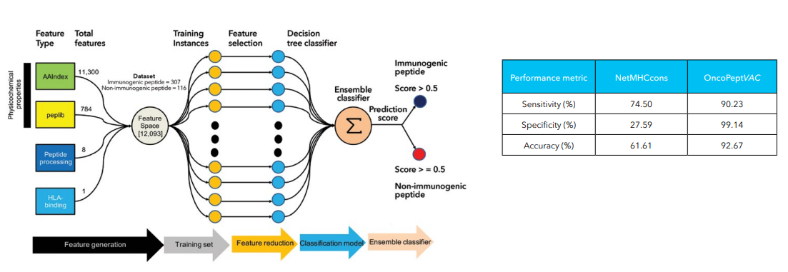
: Schematic of OncoPeptVACTM workflow and performance metrics
Figure 1: Schematic of OncoPeptVACTM workflow and performance metrics

MedGenome’s OncoPeptSCRNTM

Therapeutic revival of tumor-specific exhausted T cells using neutralizing antibodies targeting the immune checkpoint inhibitors, namely, T-lymphocyte-associated protein 4 (CTLA-4) and programmed cell death protein 1 (PD-1) has significantly improved clinical outcomes in cancer. T cells exist in a wide spectrum of functional states – from fully functional at one end of the spectrum, to fully dysfunctional at the other end. One of the factors governing the fate of the tumor response to checkpoint inhibitors is the ratio of functional to dysfunctional state of T cells, which in turn is modulated by a wide array of immune-suppressive signals present within the tumor microenvironment. Tumors that are immunologically ‘hot are characterized by high infiltration of activated T cells that also express PD1 and CTLA4. These inhibitory receptors evolved to prevent over activation of the immune system but cancer cells hijack this mechanism to their benefit by expressing the corresponding ligands driving the T cells to exhaustion.5 Targeting these checkpoint inhibitors can reverse this dysfunctional state and reinvigorate the immune response, only if they are in a ‘partially exhausted’ state. This is the basis for the development of immunotherapy drugs such as Nivolumab(anti-PD1) and Ipilimumab (anti-CTLA4) that can rescue the T cells from exhaustion.

The OncoPeptSCRNTM T-cell Activation Assays leverages the single cell transcriptomics on a 10X Genomics platform to assess immunogenicity of HLA-peptide pairs. The cancer peptides/antigens selected using our OncoPeptVACTM platform are expressed as a minigene or added from outside. These are naturally processed and presented by the T cells and subsequently sequenced using 10X single-cell RNAseq and 10X single-cell TCRseq experiments. This method was successfully used to identify T cell functional states following antigen-stimulation in an ex vivo T-cell activation system. We successfully identified functional gene clusters and molecular networks that are unique to CD8+ T cell exhausted state. The combined expression of our T cell exhaustion gene signature correlated with poor prognosis when applied to TCGA data of almost 1400 tumors from many different cancers. Furthermore, the molecular pathways identified in this study provide opportunities to develop novel therapeutic interventions specially targeting dysfunctional T cells in cancers thereby enhancing the efficacy of checkpoint inhibitors.6
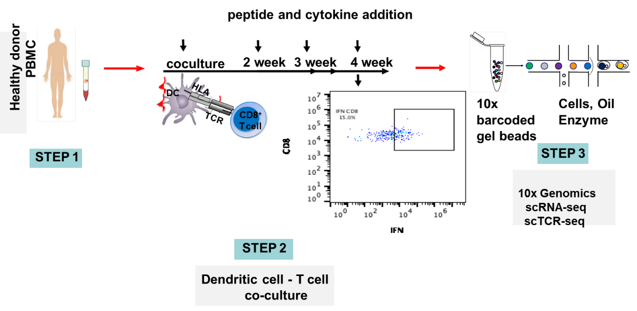
 

: Schematic of OncoPeptSCRNTM T-cell Activation Assays
Figure 2: Schematic of OncoPeptSCRNTM T-cell Activation Assays

References

  1. 1. Davda, J. et al. Immunogenicity of immunomodulatory, antibody-based, oncology therapeutics. J. Immunother. Cancer 7, 105 (2019).
  2. 2. OncoPeptVAC: A robust TCR binding algorithm to prioritize neoepitope using tumor mutation (DNAseq) and gene expression (RNAseq) data. 3 1, 223–223 (2017).
  3. 3. Majumder, S. et al. A cancer vaccine approach for personalized treatment of Lynch Syndrome. Sci. Rep. 8, 12122 (2018).
  4. 4. Mahajan, S. et al. Immunodominant T-cell epitopes from the SARS-CoV-2 spike antigen reveal robust pre-existing T-cell immunity in unexposed individuals. Sci. Rep. 11, 13164 (2021).
  5. 5. Bhojak, K. et al. Immunodominant influenza epitope GILGFVFTL engage common and divergent TCRs when presented as a 9-mer or a 15-mer peptide. http://biorxiv.org/lookup/doi/10.1101/2022.07.11.499638 (2022) doi:10.1101/2022.07.11.499638.
  6. 6. Shi, X. et al. Abstract 4943: Leveraging single-cell sequencing to discover novel exhaustion markers of CD8 T cells. Cancer Res. 79, 4943 (2019).

#T cells, #T-cell immunity, #personalized medicine, #precision therapies, #tumor mutation burden, #genomic medicine, #T-cell Activation, #anti-PD1, #anti-CTLA4, #RNA-seq data, #immune checkpoints, #genomic profiling

Immune profiling and genome sequencing solutions by MedGenome for cancer immunotherapy

According to the American Cancer Society, an estimated 1.9 million new cancers will be diagnosed in 2022 [1]. Some of the major cancer types affecting the population are prostate, lung & bronchus, colon & rectum, urinary bladder, melanoma of the skin, kidney & renal pelvis, non-Hodgkin lymphoma, oral cavity & pharynx, leukemia, pancreas, breast, colon & rectum, uterine corpus, thyroid.

By Vinay CG, Derek Vargas and Kushal Suryamohan, MedGenome Scientific Affairs

According to the American Cancer Society, an estimated 1.9 million new cancers will be diagnosed in 2022 [1]. Some of the major cancer types affecting the population are prostate, lung & bronchus, colon & rectum, urinary bladder, melanoma of the skin, kidney & renal pelvis, non-Hodgkin lymphoma, oral cavity & pharynx, leukemia, pancreas, breast, colon & rectum, uterine corpus, thyroid. Lung and Bronchus (21%) in both men and women, prostate in men (11%) and breast cancer (31%) in women are the majority cancer types causing death in the population [1]. Even though our understanding of cancer has broadened over the years it is still a major challenge to tackle across the globe. Widely accepted therapy forms for cancer includes biomarker identification and testing for treatment, chemotherapy, hormone therapy, immunotherapy, photodynamic therapy, radiation therapy, stem cell transplant, surgery, and targeted therapy. Immunotherapy (Table 1) is emerging as a forerunner among all the types of cancer therapies for the simple reason as it considers the various dynamics of immune function in an individual. Genomics has played a key role in enabling the identification of therapeutically actionable targets and in guiding the use of immunotherapy.

The magic of immunotherapy – proven right again

Recently, a team of doctors at Memorial Sloan Kettering Cancer Center published the results of a cancer trial in the New England Journal of Medicine involving Dostarlimab – a potential immunotherapy drug – to be effective in a small group of patients of 14 suffering from rectal cancer who went into complete remission [2,3]. Dostarlimab belongs to a class of drugs called checkpoint inhibitors – a programmed death 1 (PD-1) blockade drug. Mismatch Repair (MMR)-deficient colorectal cancer was found to respond well to PD-1 blockade in this trial and hence the success. PD-1 prevents T cells from killing cancer cells and thus by blocking PD-1, it is possible to activate the T-cell machinery that can then effectively kill the cancerous cells.

Mismatch repair-deficient (MMRd) or Microsatellite instability (MSI) [4] are additional factors that are linked with higher chance of developing cancer. This MMR deficiency is common in colorectal, gastrointestinal, and endometrial cancers. Finding the tumor cells with MMR deficiency can be very useful in determining the course of the treatment. The presence of MSI has also been identified as a predictor of a response to immune-checkpoint inhibition, leading the FDA to approve the anti-programmed cell death protein 1 (PD-1)-antibody pembrolizumab, for use in patients with MSI-high solid tumours regardless of histology or anatomical location.

Table 1: Types of Immunotherapies [5]

Immunotherapy Type Mode of Action
Monoclonal Antibodies (mAbs) They are the special kind of proteins which are designed to target antigens or markers present on cancer cells.
Check Point Inhibitor Drugs These drugs’ common targets are CTLA-4 and PD-1/PD-L1. The check point inhibitor drugs release the breaks allowing T cells to attack the cancer cells more efficiently.
Cancer Vaccines They trigger an immune response by identifying and attacking certain marker or antigens present on the cancer cells
Oncolytic Virus Immunotherapy Oncolytic Viruses are the genetically modified viruses that can attack the cancer cells directly. They are often combined with other types of immunotherapies such as a cancer vaccine / mAb therapy.
Adoptive T Cell Transfer It is an anti-cancer approach where the immune cells are made effective to tackle cancer. One such special approach is to add Chimeric Antigen Receptors (CARs) to T cells in the lab and reinfuse to patient. CAR T cells then can identify cancer cells and kill them.
Cytokines They aid in control and growth of immune cells
Adjuvant Immunotherapy These involve methodologies where ligands are used to boost immune response
Cancer Immunotherapy Solutions

How MedGenome’s sequencing solutions are highly effective in supporting immunotherapy?
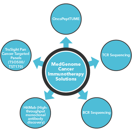
MedGenome has a primary focus on tackling immunotherapy challenges through various types of sequencing solutions:

    • OncoPeptTUME: It is a proprietary platform which interrogates RNA-Seq data sets to produce high resolution mapping of the tumor microenvironment using proprietary cell type specific gene expression signatures. It can be customized to fit cancer immunotherapy project needs and tailored to perform in preclinical and clinical settings.
    • TCR Sequencing: With the aid of Next-Generation Sequencing we offer deeper insights such as
      CDR3 repertoire diversity, clonal composition, potential antigenic recognition spectrum, and the quantity of antigen specific T-cell responses – that can be very useful in prescribing the right Immunotherapy for the patients.
    • BCR Sequencing: Our BCR sequencing solutions offer wider insights such as B-cell differentiation, BCR somatic hypermutation, class switching, and antigen specificity.
    • HitMab (High-throughput monoclonal antibody discovery): MedGenome’s HitMab platform accelerates drug discovery process through its Single Cell BCR sequencing methodologies that provide crucial information on heavy and light chain antibodies with greater specificity, enables antibody generation for low or poorly immunogenic proteins and offers distinct advantages over the current Hybridoma technology.

Our recent efforts on identifying novel neoantigens in Gallbladder Cancer published in Nature [6]:

MedGenome was part of a multi-collaborative study aimed at identifying key actionable targets and identify potential immunotherapy strategies to treat gallbladder cancers (GBC). The genomic analysis of 167 gall bladder cancer samples revealed mutated GBC genes that include several targetable driver genes such as ERBB2, ERBB3, KRAS, PIK3CA, and BRAF. Since there is no approved line of immunotherapy treatment for Gall Bladder cancers, our efforts successfully identified neoantigens from several mutated GBC genes including ELF3, ERBB2, and TP53. Validation in the lab showed T-cell activation thus indicating that they are potential cancer vaccine candidates. Additionally, some of the samples from this study had MSI which could also be targets for checkpoint inhibitor therapy.

More insights: https://cancercommunity.nature.com/posts/identification-of-actionable-targets-and-potential-immunotherapy-strategies-to-treat-gallbladder-cancers

Want to know more about our unique Cancer Immunotherapy Solutions?

Get in touch with our experienced and seasoned scientific team to understand how our unique cancer immunotherapy solutions can provide deeper insights to your research projects. You can also email us at research@medgenome.com for any queries and further details.


References

  1. 1. https://www.cancer.org/content/dam/cancer-org/research/cancer-facts-and-statistics/annual-cancer-facts-and-figures/2022/2022-cancer-facts-and-figures.pdf
  2. 3. https://www.theguardian.com/science/2022/jun/08/rectal-cancer-research-breakthrough-experimental-treatment-remission
  3. 4. https://www.mskcc.org/news/rectal-cancer-disappears-after-experimental-use-immunotherapy
  4. 5. https://www.cancer.gov/publications/dictionaries/cancer-terms/def/mismatch-repair-deficiency
  5. 5. https://www.cancerresearch.org/en-us/immunotherapy/what-is-immunotherapy
  6. 6. Pandey, A., Stawiski, E.W., Durinck, S. et al. Integrated genomic analysis reveals mutated ELF3 as a potential gallbladder cancer vaccine candidate. Nat Commun 11, 4225 (2020). https://doi.org/10.1038/s41467-020-17880-4

#Microsatellite instability (MSI), #MMR deficiency, #Immunotherapy, #CancerImmunotherapy, #Dostarlimab, #Gall Bladder cancer, #tumor mutational burden, #TCR Sequencing, #BCR Sequencing, #Checkpoint Inhibitor, #Monoclonal Antibodies, #HitMab, #Oncolytic Virus Immunotherapy, #Adoptive T Cell Transfer, #PD-1, #cancervaccine, #RNA Seq

Single Cell RNA Sequencing (scRNA-seq) – it’s role in understanding immunity and vaccine development

Next-generation sequencing techniques has seen an unimaginable growth in the past two decades. The scope has really broadened, and it is now possible to look at a genome both at macro and micro levels. Single-Cell RNA sequencing (scRNA-seq) is one such technique which deals with understanding the transcriptome at a cellular level. Single cell RNA sequencing can provide unparalleled insights into the various cellular events. scRNA-seq has an advantage over the bulk RNA-seq studies since it provides higher resolution in terms of cell subsets diversity and individual cell heterogeneity in the organisms.

By Vinay CG, Derek Vargas and Neha Varma, MedGenome Scientific Affairs

Next-generation sequencing techniques has seen an unimaginable growth in the past two decades. The scope has really broadened, and it is now possible to look at a genome both at macro and micro levels. Single-Cell RNA sequencing (scRNA-seq) is one such technique which deals with understanding the transcriptome at a cellular level. Single cell RNA sequencing can provide unparalleled insights into the various cellular events. scRNA-seq has an advantage over the bulk RNA-seq studies since it provides higher resolution in terms of cell subsets diversity and individual cell heterogeneity in the organisms.

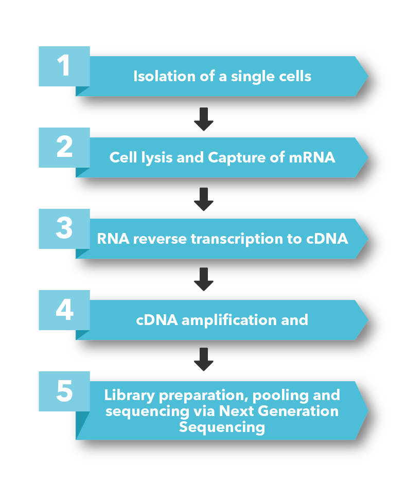
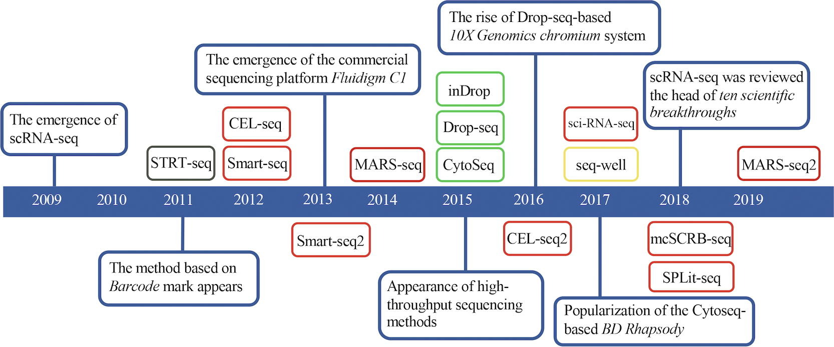
There are many scRNA-seq techniques available [1], – widely used Smart-seq2, MARS-seq, 10X Genomics, BD Rhapsody, sci-RNA-seq, in-drop and Seq-well (Figure 1) – however, all of them follow a similar approach i.e.

Library preparation, pooling and sequencing via Next Generation Sequencing


: Timeline of sc-RNA Techniques
Figure: 1 Timeline of sc-RNA Techniques
Source: https://doi.org/10.1016/j.csbj.2020.10.016

Even though scRNA-seq has several challenges one such being the high cell-to-cell variability owing to both technical and biological noise (gene expression, cellular states, cell sizes and cell cycle state) [2] – it is still an effective technique in revealing complex cellular events that can aid medical research. Now, researchers can obtain a multi-omics profiling involving genome, epigenome and transcriptome or protein information from the same single cell.

Due to its ability to provide a deeper understanding into the nature of immune cells scRNA-seq techniques can be used for:

  1. 1. Profiling the immune response to pathogens/infections
  2. 2. Evaluation of vaccines
  3. 3. Tailor personalized cancer vaccines
  4. 4. Interrogate pathogen and host transcriptomes
  5. 5. BCR analyses for mAB Development

Profiling the immune response to pathogens/infections

With scRNA-seq it is now possible to understand the molecular details of host-pathogen interaction in greater detail. Understanding the inflammatory factors triggered by immune cells in response to pathogen invasion can be useful in knowing the disease pathogenesis [3]. Few of the useful insights could be in the areas of inflammatory response analysis, identifying differently expressed genes during infection, knowing susceptible cell types, analysing infection dynamics, and studying immune repertoire [3].

Evaluation of Vaccines

scRNA-seq can be used to compare vaccine regimens and responses. Immunogenicity to vaccine is determined by several factors such as vaccine antigen, vaccine platform and adjuvant. scRNA-seq can be very useful in measuring these factors in a more specific and efficient way [1]. Besides, curating and characterizing single cell datasets of known types of pathogens at various stages of infection can help in identifying right vaccine targets [1]. scRNA-seq captures crucial information about cell types susceptible to the infection which will help the development of strategies for intervention. It can also aid in antigenic screening and selection by providing a clear insight into the immune response generated by vaccines with different antigenic makeups.

Tailor Personalised Cancer Vaccines

scRNA-seq can be extensively used in neoepitope selection and vaccine workflows. The parameters such as growth inhibition, antibody selection, cytokine secretion and a comprehensive analysis of scRNA datasets can provide deeper insights into vaccine responses. Especially, with anti-cancer drugs used to target tumor cells, some of the cells will develop resistance to such drugs. scRNA can be used to provide information on these cellular subsets that could be responsible for tumor recurrence, mutations, and pathways that can be responsible for driving tumor growth [4]. The detailed immune cell maps of multiple immunophenotypes in tumor microenvironment have been drawn via this novel sequencing method, which has deepened our understanding of tumor cell heterogeneity. These insights can be useful in identifying novel biomarkers, develop better immune responses and tailor personalized vaccines.

Interrogate pathogen and host transcriptomes

Studying the host-pathogen interaction after their encounter can provide invaluable insights into the outcomes. The scRNA-seq can help us understand which host cell types infected and which ones are able to respond to pathogens [5]. Paired dual scRNA-seq is emerging as a novel technique in understanding of those molecular pathways that would help for the host to have a distinct advantage over the pathogen. One more advantage scRNA-seq provides over the bulk studies is the high resolution it provides in identifying potential signals in rare cell populations during an infection which can help in predicting better pathogenic response, understand disease states and to identify correct biomarkers.

BCR analyses for Mab Development

scRNA-seq can provide valuable insights into full-length heavy and light chain sequences in B cells. Somatic hypermutation (SHM) and class switching are the key determinants for antibody diversity. Since, antibody discovery is important for early stage research studies, diagnostics and therapeutics, at MedGenome we have streamlined antibody discovery using high-throughput single B cell receptor sequencing and a recently licensed proprietary platform, HiTMab* (High-throughput monoclonal antibody discovery). HitMab uses high-throughput single-cell B-cell receptor sequencing (scBCR-seq) to obtain accurately paired full-length variable regions in a massively parallel fashion. Our scBCR-seq not restricted only to mice and humans but also extended to custom species including horse and rat.

Want to know more about our exciting Single-cell RNA sequencing (scRNA-seq) Solutions?

Get in touch with our experienced and seasoned scientific team to understand how our unique single-cell RNA sequencing (scRNA-seq) solutions can provide deeper insights to your research projects. You can also email us at research@medgenome.com for any queries and further details.


References

  1. 1. The Application of Single-Cell RNA Sequencing in Vaccinology, Journal of Immunology Research, Volume 2020, Article ID 8624963
  2. 2. Hedlund E, Deng Q, Single-cell RNA sequencing: Technical advancements and biological applications, Molecular Aspects of Medicine (2017), http://dx.doi.org/10.1016/j.mam.2017.07.003
  3. 3. Geyang Luo, Qian Gao, Shuye Zhang, Bo Yan: Probing infectious disease by single-cell RNA sequencing: Progresses and perspectives, Computational and Structural Biotechnology Journal, Volume 18, 2020, Pages 2962-2971, ISSN 2001-0370, https://doi.org/10.1016/j.csbj.2020.10.016.
  4. 4. Li L., Xiong F., Wang, Y. et al. What are the applications of single-cell RNA sequencing in cancer research: a systematic review. J Exp Clin Cancer Res 40, 163 (2021). https://doi.org/10.1186/s13046-021-01955-1
  5. 5. Penaranda C, Hung DT. Single-Cell RNA Sequencing to Understand Host-Pathogen Interactions. ACS Infect Dis. 2019 Mar 8;5(3):336-344. doi: 10.1021/acsinfecdis.8b00369. Epub 2019 Jan 31. PMID: 30702856.

#Single-cell RNA sequencing, #RNA sequencing, #BCR analyses, #monoclonal antibody, #cancer vaccines, #tumor, #cancer, #HitMAB, #scBCR-seq

How TCR and BCR sequencing is changing the immune research landscape

The human immune response can be divided into two components: Innate and Adaptive. Innate immune response involves classic primitive reaction through cellular and humoral mechanisms. It’s a first line of defence and can comprise a host of cells such as neutrophils, macrophages, and mast cells which kills the invading pathogens while the humoral response can be through enzymes such as Lysozyme that can kill harmful microorganisms.

By Vinay CG, Associate Director, Content & Communications and MedGenome Scientific Affairs

The human immune response can be divided into two components: Innate and Adaptive. Innate immune response involves classic primitive reaction through cellular and humoral mechanisms. It’s a first line of defence and can comprise a host of cells such as neutrophils, macrophages, and mast cells which kills the invading pathogens while the humoral response can be through enzymes such as Lysozyme that can kill harmful microorganisms.

The most effective component of the Immune system is the Adaptive immunity. Also, known as Acquired immunity – it is a highly advanced evolutionary system which recognizes, identifies pathogens, and tailors a specific response towards them. This system essentially involves Lymphocytes. Each lymphocyte expresses a receptor on its surface that can specifically bind to a particular antigen.

Even the adaptive immunity has two arms to it the cellular and the humoral. The cellular arm comprises of T Lymphocytes (T cells) which help in eliminating pathogens through various mechanisms while the humoral arm involves a subset of lymphocytes called B Lymphocytes (B cells).

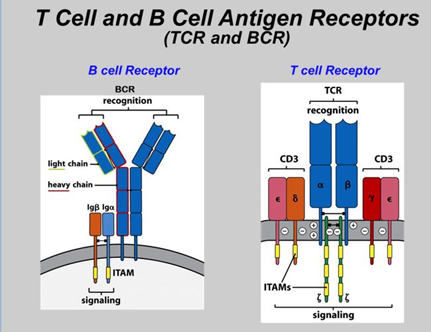
The T cells recognize antigen through T-cell antigen receptor (TCR) and the B cells recognize intact antigens through immunoglobulins (antibodies). B cells too have receptors termed as B-cell antigen receptors (BCR).

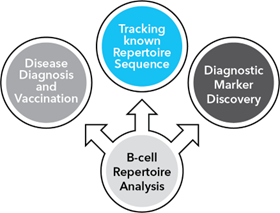
Profiling the TCRs and BCRs (Figure 1) holds a great promise in understanding mechanisms that can provide extremely useful insights in developing new therapeutics and addressing critical research questions in the areas of translational immunology, immunotherapy, autoimmune disorders, and transplant research [1,2].

TCR Profiling

TCRs are made up of heterodimers α/β (TCR2) or γ/δ (TCR1) chains [3]. These chains are encoded by 4 different gene loci namely V (variable), D (diversity), J (joining) and C (constant). A typical T cell will either express an α/β or a γ/δ receptor [4]. Similarly, BCRs are made up of heavy and light chains [4].

T cell and B cell Antigen Receptors
Figure: 1 Difference in the structure of TCR and BCR. Source: https://andrewimmunology.wordpress.com/2016/01/15/immunoglobulins-and-t-cell-receptor-differences/

The TCRs are diverse in nature owing to the extensive recombination between different V, D, and J gene segments. These rearrangements can lead to an incredibly (109-1010 sequences) large spectrum of complementary-determining region 3 (CDR3) [1]. CDR3 is critical in binding to their specific antigens. When a TCR expands by binding to an antigen there is a selective expansion in the CDR3 region of the repertoire resulting in a clonotype. With effective NGS based T cell receptor sequencing methodologies and assay it is now possible to identify all such clonotypes in a diverse repertoire of TCRs. This allows researchers to analyze the TCR repertoire in patients and aid in predicting better treatment outcomes.

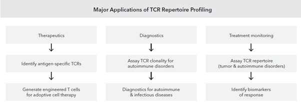
TCR repertoire sequencing can provide deeper understanding of the clonal diversity, richness and evenness of the T Cell clonal population which in turn can help in identifying biomarkers for better immunotherapy responses [5].

Major Application of TCR Repertoire Profiling

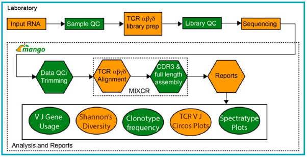
At MedGenome, we have standardized methodologies for bulk TCR sequencing where we have obtained expertise on working with diverse input types to obtain TCR α/β, γ/δ clonotypes (for bulk – cells, RNA and FFPE tissues) and TCR α/β clonotypes from single-cell inputs [1]. Additionally, our powerful bioinformatics analysis pipeline provides broader insights such as full length clonotype sequences, V-J usage summaries, CDR3 length distribution, and shared clonotype analysis (Figure 2).

Workflow for generation of bulk TCR profiling data
Figure: 2 Workflow for generation of bulk TCR profiling data.

Starting with total RNA either obtained from client or prepared in-house, sample QC is performed using the Agilent’s TapeStation or Fragment analyzer. If samples meet the QC criteria, then they are processed using the SMARTer TCR α/β Profiling kit or a modified protocol for γ/δ TCR profiling. After library preparation, QC is performed using Agilent’s TapeStation or Fragment analyzer, and sequencing is performed on Illumina platform. Data generated is demultiplexed and FastQC is performed, and after trimming, MiXCR software is used for alignment of reads to the TCR clonotypes, and the final CDR3 and full length clonotypes are assembled. Advanced analyses outputs can also be generated to compare VJ gene usage across samples, and determine Shannon’s diversity and frequency changes in the repertoire across samples.

BCR Profiling

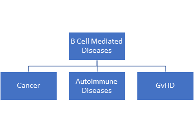
The B cells too play an important role in immune response. However, they also play havoc when they go wrong causing several B-cell mediated diseases such as cancer mostly known and noted type being B-cell malignancies such as non-Hodgkin’s lymphoma and Hodgkin’s lymphoma, autoimmune diseases such as multiple sclerosis, rheumatoid arthritis and systemic lupus erythematosus [6,7] As a B cell matures several recombination of immunoglobulin genes occur owing to an event called class switch recombination (CSR) which can lead to diversification of the B-cell repertoires [8]. Further, B-cell receptors can undergo variations when it binds to a certain antigen. through V(D)J recombination, class switch recombination, and somatic hypermutation that can cause DNA breaks triggering cancer development [8].

B-Cell Mediated Diseases

Therefore, BCR sequencing gives an insight into the various interplay between B-cell differentiation, BCR somatic hypermutation, class switching, and antigen specificity.

Major Applications of BCR Repertoire Profiling

Major Applications of BCR Repertoire Profiling

At MedGenome, we have also developed and standardized, HitMab (High-throughput antibody discovery using single cell sequencing), a streamlined workflow for antibody discovery using high-throughput single-cell B-cell receptor sequencing (scBCR-seq). HitMab allows us to obtain accurately paired full-length variable regions in a massively parallel fashion. With HitMab’s single cell BCR sequencing it is possible for researchers to speed up discovery of thousands of antibodies at a much lesser time compared to the traditional Hybridoma Technology.

Want to know more about our exciting TCR/BCR Solutions?
Get in touch with our experienced and seasoned scientific team to understand how our unique immune repertoire solutions can provide deeper insights to your research projects. You can also email us at research@medgenome.com for any queries and further details.

References

  1. 1. MedGenome TCR White Papers, https://research.medgenome.com/medgenome-documents/
  2. 2. 10x Genomics, Inc. LIT000017 Rev C Chromium Single Cell Immune Profiling Product Sheet
  3. 3. Transplant International 2019; 32: 1111–1123
  4. 4. David Male, Immunology an illustrated outline, 4th Edition, 2004, Elsevier
  5. 5. https://www.thermofisher.com/in/en/home/life-science/sequencing/sequencing-learning-center/next-generation-sequencing-information/immuno-oncology-research/why-immune-repertoire-matters.html
  6. 6. Bite-sized immunology by British Society of Immunology.
  7. 7. Bashford-Rogers, R.J.M., Bergamaschi, L., McKinney, E.F. et al. Analysis of the B cell receptor repertoire in six immune-mediated diseases. Nature 574, 122–126 (2019). https://doi.org/10.1038/s41586-019-1595-3
  8. 8. Illumina Immuno-oncology overview publication.

#t cell receptor sequencing, #tcr repertoire sequencing, #t cell repertoire sequencing, #bcr sequencing, #TCR repertoire, #NGS based t cell receptor sequencing

What’s Next for Single-Cell Genomics?

Single-cell genomic analysis has emerged as a powerful method for studying complex disease. By providing comprehensive analyses of individual cells, single-cell sequencing allows researchers to examine cellular heterogeneity, which especially useful in oncology, neurology, immunology, and developmental research.

By MedGenome Scientific Affairs

Single-cell genomic analysis has emerged as a powerful method for studying complex disease. By providing comprehensive analyses of individual cells, single-cell sequencing allows researchers to examine cellular heterogeneity, which especially useful in oncology, neurology, immunology, and developmental research.

Because it can analyze individual cells in depth, scientists can obtain unbiased cell analyses. According to Lei et al., “Single-cell sequencing significantly outperforms previous sequencing technologies in terms of our understanding of the human biology of embryonic cells, intracranial neurons, malignant tumor cells and immune cells because it can probe cellular and microenvironmental heterogeneity at single-cell resolution.”1

A relatively new technique, single-cell genomic sequencing technologies continue to improve; in turn, scientists are developing new approaches and discovering novel uses. While technological limitations remain, single-cell genomic analysis promises to advance understanding of cell types to inform novel therapies.

The next milestone in this field is single-nucleus RNA sequencing (snRNA-seq), which allowed the extension of single-cell transcriptomics analyses to human diseases for which live tissue is difficult to obtain. One of the first studies was conducted by Lake et al., which involved single-cell analysis of molecular pathology in the brain of patients with autism spectrum disorder (ASD).

What is Single-Cell Genomic Sequencing?

Scientists use single-cell genomics to study the functionality and properties of a cell.2 Whereas conventional genetic sequencing uses tissue samples to produce the average diversity of cells, single-cell genomics drills down to the single-cell level.3 This level of specificity allows scientists to study cell-to-cell variations and identify rare cells that play a role in disease progression.

Single-cell genomic techniques accomplish the following:4

  • Characterize and identify heterogeneous cell populations
  • Discover new cell markers and regulatory pathways
  • Uncover novel cell types, cell states and rare cell types
  • Reconstruct developmental hierarchies and reveal lineage relationships

Single-Cell Genomic Analysis Techniques

In its early days (a little over ten years ago), single-cell sequencing caused a stir in the scientific community, but its high cost made it impractical to use in most situations. Technology has advanced, however, enabling high-throughput single-cell sequencing via multiple profiling strategies.

In addition to single-cell RNA sequencing (RNA-seq), other sequencing platforms and methods allow scientists to capture information on cell-surface proteins, chromatin state, genetic perturbations, and genome data.5 Each method expands on RNA data to provide a unique perspective on cell state and identity.

The Single-Cell Sequencing Protocol
Preparing samples for processing and capturing individual cells is a complex process that involves four primary steps:6

  • Isolation of single cells from a cell population
  • Extraction, processing, and amplification of the genetic material of each isolated cell
  • Preparation of a “sequencing library” including the genetic material of an isolated cell
  • Sequencing of the library using a next-generation sequencer

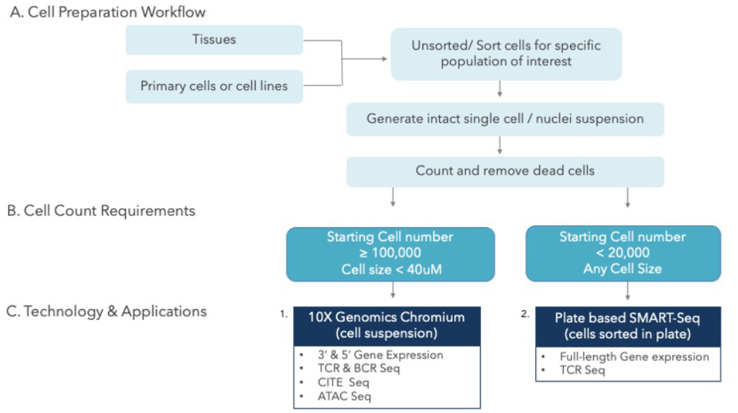
To successfully generate single-cell libraries from the diverse starting material including tissue types and cells. MedGenome has integrated several validated single-cell isolation and library preparation platforms such as Miltenyi tissue dissociation.

Single Cell Sequencing Protocol

Single-Cell Genomics Applications
Because immune cells have several distinct functions, single-cell sequencing is a valuable tool for understanding the immune system and identifying new targets for treatment.4 Researchers have used the technique to identify subpopulations of spleen and blood natural killer (NK) cells in humans and mice, contributing to translational research. Researchers have also used single-cell RNA-seq to study highly heterogeneous immune cells, helping them better understand why the immune system weakens with age.

Researchers out of Duke University recently used single-cell sequencing to study cerebral cavernous malformations (CCMs), a blood vessel abnormality that can lead to brain hemorrhage.7 Using the technique helped them answer questions about CCM pathogenesis.

Single-cell genomic techniques are becoming especially valuable to oncology researchers, allowing them to better understand treatment response and resistance, as well as inform diagnosis and monitoring.8 As a study published in Biomolecules concludes, “the molecular characteristics of each cellular component and its interconnections—either promoting or inhibiting tumor growth—are all points that can be leveraged during therapeutic development. Therefore, single-cell genomics and the related multi-omics technologies are exploratory tools that far exceed the scope and effectiveness of preceding bulk genomic analyses.” 8

Partner with a Single-Cell Genomics Solutions Expert
Whether you’re studying tumor cell response or immune cell functions, single-cell genomics is a complex but powerful method for understanding the cellular structure, behavior, and heterogeneity. Get in touch with MedGenome to explore various single-cell sequencing solutions and our abilities to design and support your experiment.

References

  1. 1 Lei, Y., Tang, R., Xu, J. et al. Applications of single-cell sequencing in cancer research: progress and perspectives. J Hematol Oncol 14, 91 (2021). https://doi.org/10.1186/s13045-021-01105-2
  2. 2 Kaur, Raman et al. Chapter 9 – Single-Cell Genomics: Technology and Applications. Single-Cell Omics, Academic Press (2019) https://doi.org/10.1016/B978-0-12-814919-5.00009-9
  3. 3 Wang Q, Yang KL, Zhang Z, et al. Characterization of Global Research Trends and Prospects on Single-Cell Sequencing Technology: Bibliometric Analysis. J Med Internet Res. 2021;23(8):e25789. doi:10.2196/25789
  4. 4 Tang X, Huang Y, Lei J, Luo H, Zhu X. The single-cell sequencing: new developments and medical applications. Cell Biosci. (2019);9:53. doi:10.1186/s13578-019-0314-y
  5. 5 Vargas, Derek. Advancing Single-Cell Multi-Omic Approaches to Biomedical Research. MedGenome blog, (2021) https://research.medgenome.com/blog/advancing-single-cell-multi-omic-approaches-biomedical-research/
  6. 6 Vaga, Stephanie Ph.D. Understanding Single-Cell Sequencing, How It Works, and Its Applications. Technology Networks, (2022) https://www.nature.com/articles/s41431-019-0508-0https://www.technologynetworks.com/genomics/articles/understanding-single-cell-sequencing-how-it-works-and-its-applications-357578#D5
  7. 7 Snellings DA, Girard R, Lightle R, et al. Developmental venous anomalies are a genetic primer for cerebral cavernous malformations. Nat Cardiovasc Res. (2022);1:246-252. doi:10.1038/s44161-022-00035-7
  8. 8 Kim N, Eum HH, Lee HO. Clinical Perspectives of Single-Cell RNA Sequencing. Biomolecules. 2021;11(8):1161. doi:10.3390/biom11081161
  9. 9 Lake BB, Ai R, Kaeser GE, Salathia NS, et al. Neuronal subtypes and diversity revealed by single-nucleus RNA sequencing of the human brain. SCIENCE 2016; 6293: 1586-1590. doi: 10.1126/science.aaf1204

#single cell genomic sequencing, #single cell sequencing, #single cell genomics, #single cell genomic analysis

Rare Disease Day 2022: A call for better Diagnosis, Treatment, and Equity

February 28th is Rare Disease Day. It is a day where the realities of Rare Diseases need to be highlighted for all health industry stakeholders; to celebrate the progress that has been made as well as to inspire us for the challenges that lay ahead.
Rare diseases are defined as those conditions thar affect fewer than 1/ 1200 people. More than 300 million people globally are affected by a rare disease 1,2. Patients and families with rare diseases are one of the most underserved communities in medicine today.

By Aditya Pai, VP Corporate and Business Development, MedGenome Inc, USA.

Rare Disease Day

February 28th is Rare Disease Day. It is a day where the realities of Rare Diseases need to be highlighted for all health industry stakeholders; to celebrate the progress that has been made as well as to inspire us for the challenges that lay ahead.

Rare diseases are defined as those conditions thar affect fewer than 1/ 1200 people. More than 300 million people globally are affected by a rare disease1,2. Patients and families with rare diseases are one of the most underserved communities in medicine today. There are 7000 + documented rare diseases, yet for the most, a cure or treatment is not available3. Only 5% of rare diseases have an approved treatment. Most rare diseases get misdiagnosed or remain undiagnosed resulting in complex testing and clinical odysseys. This is often due to the variability in symptoms from disease to disease for also for the same disease from person to person. 70% plus of rare diseases are seen in children while greater than 72% are genetic in origin.

There are three key challenges with rare disease research: 1. Diagnosis 2. Treatment 3. Equity

  1. Diagnosis: Especially in neonates, the emotional trauma caused to new parents with an undiagnosed rare disease is heartbreaking. With costs of sequencing falling, in 2022, it is possible to perform far more elaborate next generation sequencing (NGS) than even five or ten years ago. Whole genome sequencing (WGS) or whole exome (WES) / clinical exome sequencing (CES) has been proven to detect with more sensitivity and specificity previously undiagnosed rare diseases. For example, in a 2020 UK study, WGS data for 13,037 participants, of whom 9,802 had a rare disease was analyzed. A genetic diagnosis was provided to 1,138 of the 7,065 phenotyped participants. 95 Mendelian associations between genes and rare diseases were found. At least 79 of these were confirmed to be etiological. Further, a 2021 UK study for whole genome sequencing for neurological patients with repeat expansion disorders (e.g., Fragile X syndrome) showed high sensitivity and specificity, and led to the identification of neurological repeat expansion disorders in previously undiagnosed patients. The use of NGS – in particular WGS has proven to be invaluable in diagnosis.
  2. Treatment: Rare disease drug development has unique challenges. Finding patients for clinical trials, the small number of patients affected, as well as the heterogeneity of rare diseases and the way they progress makes clinical trial design and participation a challenge. Understanding the natural histories of disease can play a vital role in drug development especially when designing meaningful end points and patient inclusion / exclusion criteria. In the USA, government has provided various incentives to drug manufacturers. The results of a 2020 CDER report4 showed that of the 53 novel drugs approved in 2020, 58% were for rare diseases demonstrating the interest of pharmaceutical and biotechnology companies.
  3. Equity: Not everyone can afford whole genome sequencing or any form of genetic testing or treatments that are costly. This can lead to large inequities in diagnosis, treatment and ongoing care globally. The financial and emotional cost to families affected by rare disorders can be enormous as a result of such inequities.

A Call to Action:

  • More affordable WGS/WES/CES, its interpretation with appropriate genetic counseling and care key to stymie long and complex diagnostic odysseys.
  • Pharmaceutical companies need to continue to adopt global strategies to not only understand the natural history of specific rare diseases, but also include more countries to recruit patients for trials. For example, India and South Asia have the world largest population of people affected by rare and inherited disease. This is highly relevant, considering the large number of rare diseases and the small global pool of potential patients available for clinical trials.
  • Health policy must account for equitable access to diagnosis, treatment and care for rare disease populations. While these will vary by country, its essential to start developing a policy for rare diseases, orphan drugs and their cost-effective access.

Rare Disease Day 2022 represents another opportunity to remind ourselves of the work that remains. At MedGenome, we have formed a strategic alliance with Emmes across six rare disorders with the goal of expanding and accelerating Rare Disease research and development. Our partnership will combine patients’ genomic, phenotypic and epidemiological data into custom rare disease registries. MedGenome in India routinely offers CES and has solved many undiagnosed conditions successfully. Through our clinical collaborators, we continue to highlight the unique aspects of Indian genomes in the understanding the natural history and heterogeneity of rare diseases as well as the role they can play in global rare disease clinical trials.

References

  1. 1 Am J Med Genet A. 2019;179(6):885–892. http://doi.org/10.1002/ajmg.a.61124.
  2. 2 Eur J Hum Genet. 2020;28(2):165–173. https://www.nature.com/articles/s41431-019-0508-0
  3. 3 Ibid.
  4. 4 https://www.fda.gov/media/144982/download

#Rarediseases, #Rare disease research, #diagnosis, #treatment, #equity, #next-generation sequencing #Whole genome sequencing #clinical trial design

Transform your cancer research with the most suitable “omics” strategy

World Cancer Day is a day to reflect and celebrate research victories, the battles that anyone with cancer fights, the search for new ways to detect cancer early and treat it as effectively as possible. Yet, cancer statistics remain sobering. Globally, there were an estimated 19.3 million new cancer cases and 10 million cancer deaths in 2020 . The number of people living with cancer is expected to grow by around 1 million every decade between 2010 and 2030.

By Aditya Pai, VP Corporate and Business Development, MedGenome Inc.

World Cancer Day is a day to reflect and celebrate research victories, the battles that anyone with cancer fights, the search for new ways to detect cancer early and treat it as effectively as possible. Yet, cancer statistics remain sobering. Globally, there were an estimated 19.3 million new cancer cases and 10 million cancer deaths in 2020i. The number of people living with cancer is expected to grow by around 1 million every decade between 2010 and 2030ii.

Over the past decade, a better understanding of alterations in single driver genes, and genes involved in specific biological pathways have led to the development of better targeted therapies and more recently, immunotherapies. For example, tyrosine kinase inhibitors for non-small cell lung cancer or checkpoint inhibitors used as tissue agnostic immunotherapies have altered the treatment of many cancers. Yet, understanding the molecular mechanisms of various cancers is critical to develop new screening methods, e.g. non-invasive circulating tumor DNA ctDNA tests and novel therapies. The quest to improve overall survival rate in cancer with better and more targeted treatments has been abetted by significant investments by pharmaceutical and device companies as well as government funding. Precision medicine has also been aided by decreased costs of sequencing the genome which at large scale was cost prohibitive in the past. This has led to complex choices and decisions around whether to use a targeted gene panel or sequence the exome or whole genome. Various “omics” technologies have led to understanding the changes at the DNA level in a tumor to understanding the expression of various changes at the level of the transcriptome to understanding the tumor microenvironment using single-cell sequencing approaches. This multi-omic paradigm is gaining rapid traction in research. Yet, for a translational oncology researcher, a question I often get asked is “Should I sequence the entire genome or use a combination of whole exome sequencing and DNA sequencing or should I use a cancer panel?”

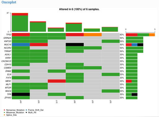
A translational oncology researcher is often looking for a broad set of results from a retrospective set of tumor samples stored as FFPE blocks (Formalin Fixed Paraffin Embedded). In this research discovery scenario, either of the above approaches have value, but it depends on factors such as the research hypothesis, time, research budget as well bioinformatics capabilities. For example, a Research Use Only (RUO) comprehensive gene panel like TSO 500 from Illumina includes 523 cancer related genes, includes (1) DNA variations like SNV’s, indels, copy number alterations (2) RNA gene fusion detection and (3) broad biomarker measurements like TMB that integrate many genomic loci across the genome. The TSO 500 panel has the flexibility in being used with solid tumor or ctDNA. The panel comes with added benefits of the bioinformatics pipeline included in the workflow. With more advanced bioinformatics, as we provide at MedGenome (see Figure 1, 2), a researcher could explore various hypothesis to better understand potential pathways and upstream and downstream cancer driver genes. Panels like TSO 500 are rigorously developed and designed to provide very robust, specific and sensitive (typically down to 5% variant allele frequency for most applications) detection of alterations in the genes of interest.

This same researcher could use a whole exome and RNA sequencing or whole genome sequencing approach. However, unlike the TSO 500 assay where bioinformatics analysis is included in the workflow, the researcher would have to separately perform bioinformatics analysis that can be time consuming. Yet, they may find this worthwhile if they are needing to explore the genome beyond what a panel of 523 genes would cover.

Key Takeaway
Sequencing and multi-omic approaches in oncology are quickly evolving and must be tailored to the primary questions that an oncology researcher is trying to answer. There are many approaches available.

At MedGenome, we can help you with your “omics” strategy. These include panel-based approaches that leverage comprehensive cancer genes, as well, whole genome / whole exome-based approaches, or more comprehensively single-cell RNA sequencing approaches.

In all these choices, cost is also an important consideration as is the ultimate use of these results which can range from exploratory research to a specific path for companion diagnostic development for a drug in clinical trials. Appropriate effort and detail to study design of a sequencing strategy and method is critical, as is the need for collaboration with high quality laboratories with validated assays.

Oncoplot displaying the somatic landscape

Figure 1: Oncoplot displaying the somatic landscape of the cohort for top (max 20) most frequently mutated genes. Each row represents a gene and each column represents a sample. Colored squares show mutated genes, while grey squares show non-mutated genes

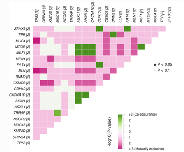
Somatic interaction plot

Figure 2: Somatic interaction plot shows mutually exclusive or co-occurring pair of genes displayed as a triangular matrix with top (max 20) most frequently mutated genes in the cohort. Green indicates tendency toward co-occurrence, whereas pink indicates tendency toward exclusiveness.

References

  1. I. https://www.uicc.org/news/globocan-2020-new-global-cancer-data
  2. II. https://www.macmillan.org.uk/_images/people-living-with-cancer_tcm9-283689.pdf


#cancer research, #targeted therapies, #tyrosine kinase inhibitors, #immunotherapies, #non-small cell lung cancer, #checkpoint inhibitors, #targeted gene panel, #translational oncology, #multi-omic approaches, #single-cell RNA sequencing

Spatial Transcriptomics: Beyond gene expression via tissue architecture

Spatial transcriptomics is a revolutionary molecular profiling method that allows scientists to measure in a tissue sample and map the activity to specific cell types and their location. This novel technology is paving the path to new discoveries that are proving instrumental in helping researchers gain a better understanding of biological processes and diseases leading it to be called the Method of the Year in 2020.

By Dr. Neha Verma, Research Scientist – NGS, MedGenome Inc., USA

Spatial transcriptomics is a revolutionary molecular profiling method that allows scientists to measure in a tissue sample and map the activity to specific cell types and their location. This novel technology is paving the path to new discoveries that are proving instrumental in helping researchers gain a better understanding of biological processes and diseases leading it to be called the Method of the Year in 2020.

The beginnings of spatial transcriptomics can be traced back to the 1960s where nucleic acids were stained at their original locations with cells or tissues. Although the term spatial transcriptomics was first coined in 2016, the first steps were already taken in the late ’60s with the use of in situ hybridization. This was followed in the late ’90s by the first microdissection techniques, in which a microscope is used to dissect a small portion of tissue. The term “Spatial Transcriptomics” is a variation of Spatial Genomics, first described by Doyle, et. al., in 2000 and was then modified by Ståhl et. al. in 2016.

Single cell/nuclei sequencing plays a crucial role in identification of cellular subpopulations and their response to various conditions/stimuli. Cells are impacted by their native environment and surroundings. Understanding cellular responses in their endogenous spatial context. With spatial transcriptomics, it is now possible to obtain information on the transcriptomes of a single cell or a small group of cells, while maintaining the information on where the cell (or group of cells) is located within the tissue.

Currently, there are only a few different types of spatial transcriptomics techniques that are available. These include GeoMx from NanoString; Slide-seq, Apex-seq; High-Definition Spatial Transcriptomics (HDST); and 10X Genomics’ Visium Spatial Gene Expression.

GeoMx
NanoString’s GeoMx Digital Spatial Profiler allows to define a microscopic region of interest on an FFPE or frozen tissue slide due to a UV-photocleavable barcode engineered into the in-situ hybridization probes. The region of interest is specifically exposed to UV light, and the barcodes are cleaved, used to identify the RNA or protein present in the tissue. The size of the defined regions of interest can vary in between ten to six hundred micrometers allowing targeting of a wide variety of structures and cells in the histological sample.

APEX-seq
The method utilizes the APEX2 gene, expressed in live cells which are incubated with biotin-phenol and hydrogen peroxide. In these conditions, the APEX2 enzymes catalyse the transfer of biotin groups to the RNA molecules, and these can then be purified via streptavidin bead purification. The purified transcripts are then sequenced to determine which molecules were near the biotin tagging enzyme.

Slide-seq
Slide-seq relies on the attachment of RNA binding, DNA-barcoded micro beads to a rubber-coated glass coverslip. The microbeads are mapped to their spatial location via SOLiD sequencing. Tissue sections are transferred to this coverslip to capture extracted RNA. Captured RNA is amplified and sequenced. Transcript localization is determined by the barcode oligonucleotide sequence from the bead that captured it.

High-Definition Spatial Transcriptomics (HDST)
It is based on decoding the location of mRNA capture beads in wells on a glass slide. This is accomplished by sequential hybridization to the barcode oligonucleotide sequence of each bead. Once the location of each bead is decoded, a tissue sample can be placed on the slide and permeabilized. The captured transcripts are then sequenced. HDST uses smaller beads than Slide-seq and thus can resolve at a spatial resolution of two micrometers compared to ten micrometers of Slide-seq.

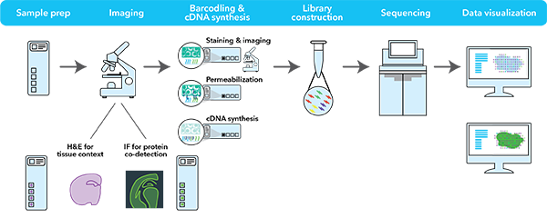
And the very latest breakthrough by 10X Genomics, Visium Assay. The Visium spatial assay combines traditional histopathology with unbiased, high-throughput gene expression analysis from the same tissue section at high resolution and sensitivity. This enables spatial clustering of cells based on gene expression that reliably correlates with the neuroanatomy of intact tissue, across different mammalian brain regions. The addition of immunofluorescence staining enables the simultaneous examination of protein and gene expression from the same tissue, providing additional insights.

10X Genomics

Source: 10X Genomics

The 10X Visium assay is a newer and improved version of the Spatial Transcriptomics assay which also utilizes spotted arrays of mRNA-capturing probes on the surface of glass slides but with increased spot number, minimized spot size and increased amount of capture probes per spot. Within each of the four capture areas of the Visium Spatial Gene Expression slides, there are approximately 5000 barcoded spots, which in turn contain millions of spatially barcoded capture oligonucleotides. Tissue mRNA is released upon permeabilization and binds to the barcoded oligos, enabling capture of gene expression information. Each barcoded spot is 55 µm in diameter, and the distance from the center of one spot to the center of another is approximately 100 µm. The spots are staggered to minimize the distance between them. On average, mRNA from anywhere between 1 and 10 cells are captured per spot which provides near single-cell resolution. Each Visium Spatial Gene Expression Slide includes 4 capture areas (6.5 x 6.5 mm), each defined by a fiducial frame (fiducial frame + capture area is 8 x 8 mm). The capture area has ~5,000 gene expression spots, each spot is ~55 microns with primers that include:

Illumina TruSeq Read 1 (partial read 1 sequencing primer). 16 nt Spatial Barcode (all primers in a specific spot share the same Spatial Barcode); 12 nt unique molecular identifier (UMI); 30 nt poly(dT) sequence (captures poly-adenylated mRNA for cDNA synthesis). Distance from center to center of each spot is ~100 microns.

Tissue sections on the capture areas of the Visium Spatial Gene Expression Slide are fixed using methanol. Hematoxylin is used to stain the nuclei, followed by eosin staining for the extracellular matrix and cytoplasm. The stained tissue sections are imaged. The same tissue section is permeabilized to release mRNA onto capture spots that contain spatially barcoded oligos fixed to the slide. mRNAs are converted to cDNAs and then collected for dual-indexed Illumina library construction and sequencing. The H&E stained image and the spatially barcoded cDNAs are overlaid to allow visualization of the gene expression within the original tissue placement.

The tissue sections should be no larger than the capture area (6.5 mm x 6.5 mm) to avoid covering the fiducial frame that is used to align the RNASeq data with the stained tissue images. In addition, tissue placed outside the capture area will also simply not generate any additional gene expression data, or could possibly convolute the gene expression data generated.

With Visium’s whole transcriptome and protein co-detection approach

  • Gain insights on cell-to-cell interactions with spatial context: Discover new biomarkers by examining histology, protein, and mRNA from the same fresh frozen tissue section
  • Characterize cellular sub-types and functional states: Reveal the spatial organization of newly discovered cell types, states, and biomarkers with whole transcriptome analysis
  • Discover regional cell heterogeneity throughout: Examine gene and protein expression heterogeneity and how it contributes to biological system

Spatially resolved gene expression can provide a powerful complement to traditional histopathology methods, enabling a greater understanding of cellular heterogeneity and organization within the tissue architecture.

References

  1. Rao A, et al. Exploring tissue architecture using spatial transcriptomics. Nature. 2021 Aug; 596(7871): 211–220.
  2. Application Note: Enriching pathological analysis of FFPE tumor samples with spatial transcriptomics. LIT000152 – Rev A, https://pages.10xgenomics.com/rs/446-PBO-704/images/LIT000152_ApplicationNote_Visium_FFPE_Prostate_Cancer.pdf
  3. Ståhl PL, et al. Visualization and analysis of gene expression in tissue sections by spatial transcriptomics. Science 353: 78–82, 2016. doi: 10.1126/science.aaf2403


#Spatial Transcriptomics, #Spatial gene expression, #Spatial Genomics, #Transcriptomes, #Cellular heterogeneity

Impact and applications of NGS: Opening the doors into the world of “omics”

It is known that all the hereditary information is contained within an organism’s genome. Owing to continuous global efforts many new bioinformatics databases are emerging and has seen an up trend in the recent past, a reflection on how NGS data is impacting our understanding of life and our need to constantly develop new methods to investigate and decode the information in and around DNA (or RNA for some viruses) and its nucleotide sequences.

By Parimala Nagaraja, Assistant Manager-NGS, MedGenome Inc., USA

It is known that all the hereditary information is contained within an organism’s genome. Owing to continuous global efforts many new bioinformatics databases are emerging and has seen an up trend in the recent past, a reflection on how NGS data is impacting our understanding of life and our need to constantly develop new methods to investigate and decode the information in and around DNA (or RNA for some viruses) and its nucleotide sequences.

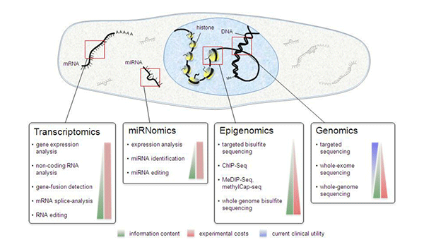
NGS Sequencing Types
Fig: 1 Applications of NGS in different Areas of Biological research.

Source: Next-Generation Sequencing: From Understanding Biology to Personalized Medicine – Scientific Figure on ResearchGate. Available from: https://www.researchgate.net/figure/Next-generation-sequencing-applications-Schematogram-depicting-the-different-methods-for_fig1_262384938 [accessed 15 Nov, 2021]

Genomics:

A comprehensive outlook and understanding of a full genome is now possible with de novo whole-genome shotgun sequencing and annotation. Because of the novel technological developments over the recent years and the availability of several reference genomes in the public domain that can be used for annotation, WGS has become increasingly easier, faster, and cheaper. NGS plays a significant role in Human genomics ushering a new era of new personalized therapeutics in order to achieve healthy and disease-free lifestyle – the broad term being“personomics”. Several SNPs, mutations, other sequence variants such as InDels, copy number (CNVs), and structural variations (SNVs) can be detected through Targeted sequencing like Ampliseq or Whole Exome Sequencing (WES) within or among different species. Other methods of Targeted sequencing that are widely used for identifying polymorphisms that are important in tissue or cell matching for transplantation include HLA genotyping of an entire gene or just exonic regions. Targeted sequencing of just coding regions to detect exonic mutations responsible for rare Mendelian Genetic disorders such as hearing loss, intellectual disabilities, and movement disorders and for investigating common disorders such as heart disease, hypertension, diabetes, and cancer can be termed as “Exomics”.

Transcriptomics:

The sum total of RNA transcript sets expressed by the genome in cells, tissues, and organs at different stages of an organism’s life cycle is termed as a “transcriptome” of an organism. High throughput RNA sequencing from Complementary DNA (cDNA) molecules helps us to understand the complex and intricate genome functions in biological systems. It also provides us to identify quantitative expression levels of genes, tissue specific transcript variants and isoforms, small and large non-coding RNAs involved in the regulation of gene expression or associated with various types of cancer in a highly sensitive and accurate manner.

Methylomics and epigenomics:

The study of complete epigenetic modifications via DNA nucleotide methylation and posttranslational modifications of histones, the interaction between transcription factors and their targets, and nucleosome positioning is called Epigenomics. The genome-wide analysis (GWA) of DNA methylations and their effects on gene expression and heredity is called Methylomics. Bisulfite DNA sequencing (Methyl-seq) aids in mapping DNA cytosine methylation at single-base resolution. Methyl seq is a well-established method for DNA methylation profiling in various organisms as well as humans for evaluating pathogenic variants of the genes.

ChIP-seq (Chromatin Immunoprecipitation) allows the genome-wide profiling of DNA-binding proteins and histone and nucleosome modifications. It is the most widely used method for detecting and analysing the transcription factor binding sites and histone modifications in a variety of organisms. Another commonly used NGS method used in epigenomics is Hi-C which is generally used to identify DNA regions such as promoters, enhancers, and insulators that come together to mediate their regulatory activities.

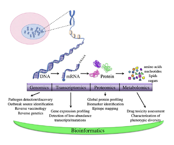
Proteomics, metabolomics, and systeomics:

Proteomics is the study of structure, function and characterization of different peptides and proteins. Sequencing the Open reading frames (ORFs) of the genomic regions, exonic regions, and transcripts aids in constructing proteomic profiles from NGS data. Although this is not the only method to build the proteomics data. A variety of several other hardware and software tools are employed to build up an organism’s peptide and protein profiles. These include 2D-PAGE, liquid chromatography coupled with tandem mass spectrometry, affinity-tagged proteins, and yeast two-hybrid assays.

The study of an organism’s total metabolic response to an environmental stimulus or a genetic modification is called Metabolomics. The metabolomics of an organism is mainly drawn from the known functions of enzymes and proteins involved in metabolic and biochemical pathways. This field forms an integral part of functional genomics in determining the phenotypic effects of genetic modifications such as gene deletions, insertions, and other mutations.

Integration of genomics, proteomics, metabolomics into a single network system is termed as Systeomics. This field of study uses computational techniques to analyse and model cell interactions. This is an interdisciplinary field of study that focuses on complex interactions within biological systems using a holistic approach.

Metagenomics:

The study of the total genomic content of the microbial community is called Metagenomics. It helps in epidemiological study of various pathogenic agents such as mycobacteria, S. aureus, E. coli, cholera, influenza, HIV, Ebola virus, etc. The Earth microgenome project reconstructed approximately 500 million varieties of microbial genomes. Before the first NGS platforms emerged, metagenomics studies were focused on 16srRNA genes to genotype and detect different species of microbes. Over the past 10 years many big projects such as the TerraGenome project for soils and the Tara Oceans project on the microbiome, eukaryotic plankton, and viromes of the global oceans emerged for sequencing metagenomes.

Agrigenomics

Studies involved in advancing crop improvements and understanding plant biology using NGS are called Agricultural genomics or Agrigenomics. Arabidopsis thaliana was the first plant genome that was published in 2000. Since then, nearly 54 new plant genomes have been sequenced in 2013, followed by another 6 plant genomes including the hexaploid bread wheat genome.

Peek into the near future:

NGS is the science of Biological information systems and “Big Data” today. However, several challenges still persist with regard to NGS data acquisition, storage, analysis, integration, and interpretation. Hence, future developments will unquestionably depend on new technologies and large-scale collaborative efforts from multidisciplinary and international teams to continue generating comprehensive, high-throughput data production and analysis. With the new innovations and the availability of cost-effective sequencing methods and the existing “Third generation sequencing” tools, smaller industries and individual scientists will be able to participate in the genomics revolution and contribute new knowledge to the different fields of structural and functional “Omics” in the life sciences.

Downstream Applications of NGS
Fig: 2. Downstream Applications of NGS in different field of “Omics”

Source: Translational research in infectious disease: Current paradigms and challenges ahead – Scientific Figure on ResearchGate. Available from: https://www.researchgate.net/figure/A-top-down-explanation-of-omics-Genomics-is-the-study-of-the-complete-set-of_fig1_225062532 [accessed 15 Nov, 2021]


References

Next-Generation Sequencing — An Overview of the History, Tools, and “Omic” Applications | InTechOpen, Published on: 2016-01-14. Authors: Jerzy K. Kulski


#NGS data, #Next-Generation Sequencing, #de novo whole-genome, SNPs, Ampliseq, Whole Exome Sequencing, Mendelian Genetic disorders, #RNA transcript, #RNA sequencing, #Methylomics, #Proteomics, #Metabolomics, #Systeomics, #Metagenomics, #Agrigenomics,