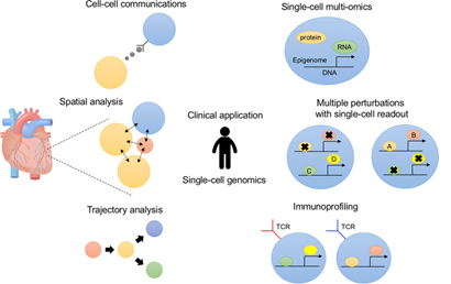
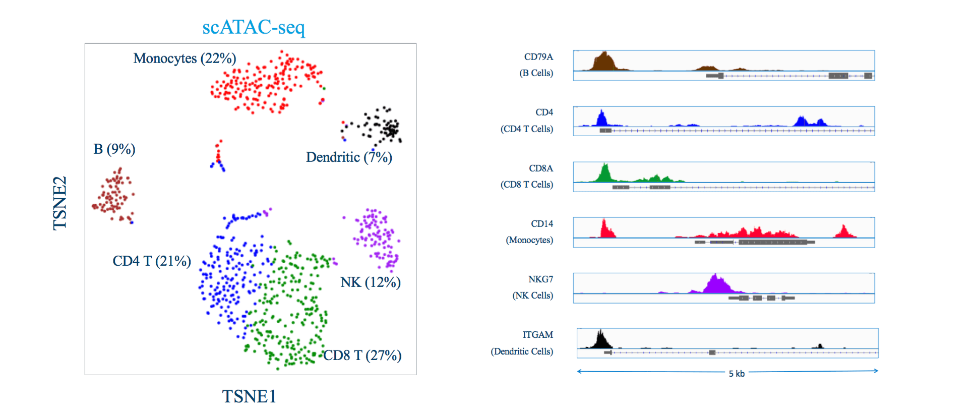
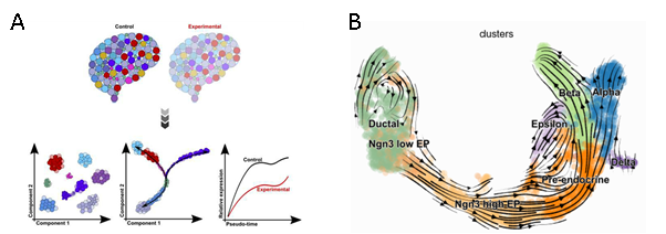
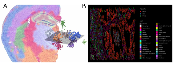
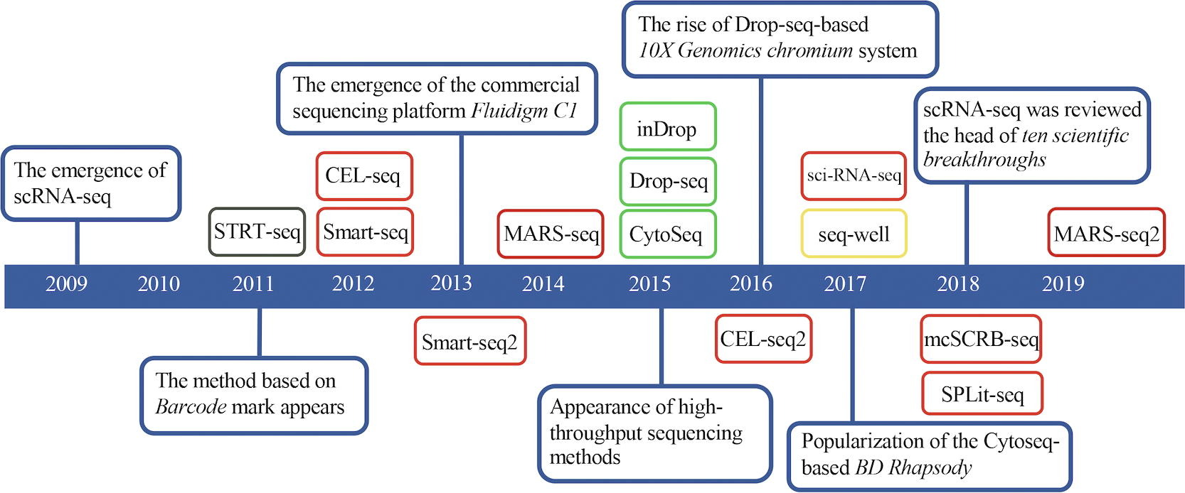
Exploring the new Human Cell Atlas release and its impact on genomic research

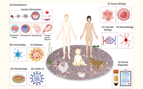
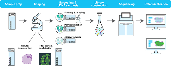
The Human Cell Atlas (HCA) consortium has unveiled a landmark collection of over 40 studies, marking a significant leap in our understanding of the human body. Published across Nature and its affiliated journals, this release represents years of collaborative effort, utilizing cutting-edge single-cell and spatial transcriptomics technologies to map the cellular composition of tissues in unprecedented detail.

By Courtney Nirenberchik, Marketing Manager, MedGenome Inc

The Human Cell Atlas (HCA) consortium has unveiled a landmark collection of over 40 studies from more than 100 countries, marking a significant leap in our understanding of the human body. Published across Nature and its affiliated journals, this updated release represents years of collaborative effort, utilizing cutting-edge single-cell and spatial transcriptomics technologies to map the cellular composition of tissues in unprecedented detail. These findings pave the way for breakthroughs in regenerative medicine, diagnostics, and the treatment of complex diseases.

Key insights from the HCA studies

The studies span diverse biological systems and disease contexts, shedding light on human development, immune responses, and tissue-specific functions. Highlights include:

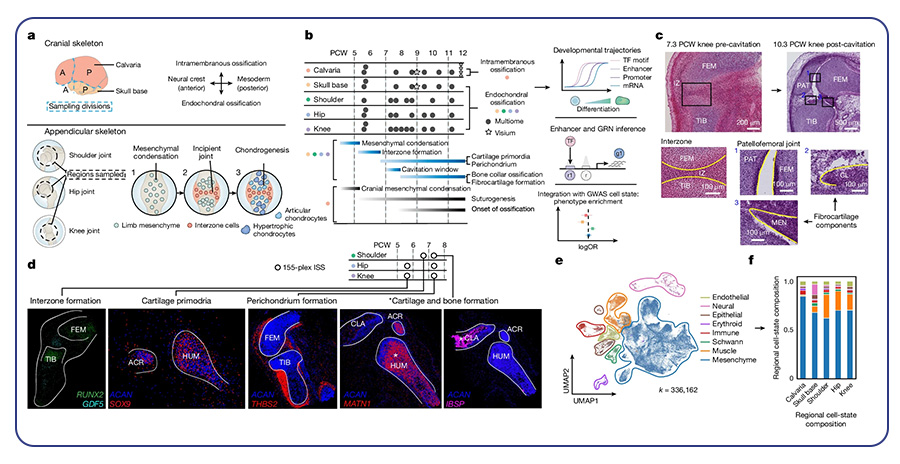
  • Developmental Biology: An atlas of the skeletal system’s formation during embryonic development offers insights into bone growth and disorders​.
  • Gut Health: A detailed map of the gastrointestinal system identified novel cell types implicated in inflammatory diseases like Crohn’s disease, providing potential targets for therapeutic intervention.
  • Immune System Insights: Comprehensive profiling of immune cells across tissues reveals new aspects of their function in maintaining health and responding to disease.

These studies not only enrich our understanding of human biology but also lay the groundwork for precision medicine by providing detailed references for healthy tissues.

multi-omic atlas of human embryonic skeletal development
To, K., Fei, L., Pett, J. P., Roberts, K., Blain, R., Polański, K., Li, T., Yayon, N., He, P., Xu, C., Cranley, J., Moy, M., Li, R., Kanemaru, K., Huang, N., Megas, S., Richardson, L., Kapuge, R., Perera, S., . . . Teichmann, S. A. (2024). A multi-omic atlas of human embryonic skeletal development. Nature, 635(8039), 657-667. https://doi.org/10.1038/s41586-024-08189-z

Advancing research with the Human Cell Atlas

The HCA’s datasets are already transforming research by serving as a gold-standard reference for single-cell and spatial transcriptomics studies. Researchers can now compare their findings against these comprehensive atlases to identify deviations associated with diseases. Moreover, the tools and methodologies developed as part of the HCA initiative are accelerating discoveries across biomedical research.

Dr. Marco Corbo, PhD, MedGenome’s Senior Scientist and Director of Bioinformatics, notes:

“The latest release from the Human Cell Atlas provides an invaluable reference for understanding the cellular basis of health and disease. By incorporating these datasets into our analyses, we can offer our clients more precise cell type annotations and deeper insights into tissue microenvironments, ultimately accelerating their research and discovery efforts.”

Empowering MedGenome’s clients with upgraded offerings

At MedGenome, we are committed to staying at the forefront of genomic research, and the latest Human Cell Atlas release directly enriches the services we provide to our clients. By integrating these groundbreaking datasets into our single-cell and spatial analysis offerings, we can deliver:

  • Enhanced Cell Type Annotation: Leveraging the HCA’s robust references, we provide highly precise annotations, ensuring our clients receive the most accurate insights from their data.
  • Improved Insights into Tissue Microenvironments: With access to this comprehensive data, our spatial transcriptomics services offer deeper understanding of cell-cell interactions and tissue-level dynamics.
  • Upgraded Tools and Reports: We are refining our bioinformatics pipelines and data visualization tools to reflect the latest methodologies and standards established by the HCA.
  • Future-Ready Research: As the HCA grows, we are poised to integrate new datasets and insights, ensuring that our clients’ research benefits from the most up-to-date scientific knowledge.

These advancements underscore our commitment to delivering services that empower researchers to push the boundaries of discovery in oncology, immunology, rare diseases, and more. The Human Cell Atlas is not just a scientific milestone—it is a resource that drives real-world applications and fuels innovation for our clients.

Curious about the analyses we offer as part of the MedGenome comprehensive solutions for understanding biology? Check out our blog posts on both single-cell and spatial analysis and explore our limited-time offer for discounted single-cell and spatial transcriptomics projects.


#Human Cell Atlas, #Developmental Biology, #spatial transcriptomics, #single-cell, #spatial analysis


Lung Cancer: Molecular Insights and Emerging Therapeutic Approaches

Lung cancer remains the leading cause of cancer-related deaths globally, accounting for 18.4% of all cancer fatalities. Smoking is the primary cause of lung cancer, responsible for 80-90% of lung cancer deaths, though other risk factors like secondhand smoke, radon, asbestos, and family history also play roles.

By Dr. Lavanya Balakrishnan and Vinay C. G., MedGenome Scientific Affairs

Lung cancer remains the leading cause of cancer-related deaths globally, accounting for 18.4% of all cancer fatalities. Smoking is the primary cause of lung cancer, responsible for 80-90% of lung cancer deaths, though other risk factors like secondhand smoke, radon, asbestos, and family history also play roles. Research efforts to improve early detection, such as low-dose CT scans and biomarker tests, offer promise, and machine learning tools are being developed to enhance CT scan accuracy1,2.

Classification and subtypes of lung cancer

Lung cancer is categorized into two main types: non-small cell lung cancer (NSCLC) and small cell lung cancer (SCLC). NSCLC accounts for 80-85% of cases and includes subtypes like adenocarcinoma (LUAD), squamous cell carcinoma (LUSC), and large cell carcinoma (LCC). LUAD, the most common subtype, originates from mucus-producing lung cells and disproportionately affects non-smokers, especially women, constituting about 40% of NSCLC cases. LUSC, which makes up 20-30% of NSCLC, is strongly associated with smoking and tends to exhibit distinct mutation profiles. LCC, though less common (9-10%), is aggressive and poorly differentiated, often presenting as a large mass. SCLC is highly aggressive, with a five-year survival rate of only 5% among heavy smokers2.

Genetic drivers of lung cancer progression

Lung cancer is driven by diverse genetic alterations that affect its development, progression, and treatment response. In NSCLC, common driver mutations include EGFR, KRAS, MET, BRAF, ALK, ROS1, and RET. LUAD is frequently associated with mutations in tumor suppressor genes like TP53, STK11, and KEAP1, while LUSC tends to involve mutations in FGFR1 and AKT1. In SCLC, aggressive growth is driven by mutations in tumor suppressors such as TP53 and RB1, alongside the activation of MYC oncogenes, leading to rapid tumor progression and treatment resistance3.

Immunotherapy: Transforming lung cancer treatment

Lung cancer has traditionally been difficult to treat due to its high mutational burden and rapid progression, which often limit the effectiveness of conventional therapies like surgery, chemotherapy, and radiation. Targeted therapies have improved outcomes for some NSCLC cases with specific genetic mutations, but most individuals, including those with SCLC, still rely on platinum-based chemotherapy, which provides limited benefit.

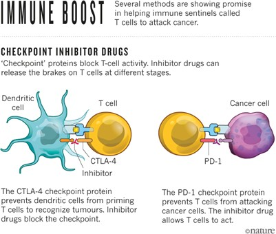
Among the various immune checkpoints that tumors exploit to evade the host immune system, the most well-known and clinically advanced are the programmed cell death protein-1 (PD-1)/programmed cell death ligand-1 (PD-L1) and cytotoxic T-lymphocyte antigen-4 (CTLA-4) pathways. The introduction of immune checkpoint inhibitors (ICIs) has revolutionized lung cancer treatment, particularly in NSCLC. ICIs like nivolumab, pembrolizumab, and atezolizumab block PD-1 and PD-L1, proteins that typically prevent T-cells from attacking cells. By releasing these “brakes,” ICIs empower T-cells to target and destroy cancer cells. Clinical trials have demonstrated the efficacy of these treatments. For instance, the KEYNOTE-024 trial, a landmark study, showed that pembrolizumab significantly improved overall survival in individuals with advanced NSCLC and high PD-L1 expression compared to standard chemotherapy. Additionally, the IMpower150 trial revealed that combining atezolizumab with chemotherapy and bevacizumab improved survival in advanced NSCLC cases, including those with lower PD-L1 expression. These breakthroughs have transformed the treatment landscape, offering substantial survival benefits, particularly in NSCLC cases without specific actionable mutations, though the impact in SCLC has been more modest, with only slight improvements when ICIs are combined with chemotherapy4.

Role of biomarkers in immunotherapy response

Although immunotherapy has transformed the treatment landscape, not all individuals respond to ICIs, and some experience severe immune-related side effects. Biomarkers such as PD-L1 expression and tumor mutational burden (TMB) can help identify those who may respond favorably to immunotherapy. For instance, higher PD-L1 expression is often associated with better outcomes in metastatic NSCLC, while elevated TMB has been linked to improved responses in certain lung cancer subtypes. However, co-mutations, such as those involving STK11/LKB1, can predict poor outcomes, and individuals with EGFR mutations or ALK fusions generally show low response rates to ICIs5.

Circulating tumor DNA (ctDNA) has emerged as a promising biomarker for monitoring response to immunotherapy, though further validation is needed to establish its clinical utility. Recently, researchers have identified a set of 140 genes that may predict better outcomes in individuals with NSCLC undergoing immunotherapy combined with low-dose radiation, potentially offering new avenues for personalized treatment strategies6.

Single-cell sequencing in lung cancer immunotherapy

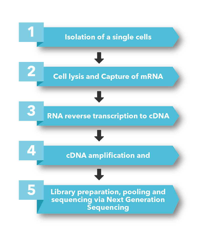
Single-cell sequencing has emerged as a pivotal tool for unraveling immunotherapy sensitivity in lung cancer. This advanced technology enables researchers to investigate the genetic, cellular, and molecular landscapes of tumors at a granular level, particularly in the tumor microenvironment (TME). By studying how different cells, including immune cells, interact within tumors, single-cell sequencing has uncovered mechanisms of immune regulation and resistance to therapies, especially immune checkpoint inhibitors (ICIs).

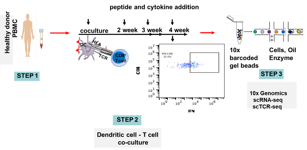
Research using single-cell RNA sequencing (scRNA-seq) has identified key immune cell populations in both non-small cell lung cancer (NSCLC) and small cell lung cancer (SCLC), shedding light on the TME’s role in patient response to immunotherapy. Importantly, studies have linked low immune cell infiltration in the TME to better responses to ICIs, illustrating the impact of tumor heterogeneity on treatment resistance. Multi-regional sampling and sequencing have further mapped clonal evolution in lung tumors, helping predict resistance and improve treatment strategies7.

Additionally, single-cell sequencing has identified new biomarkers and therapeutic targets in lung cancer. For instance, KAT2B has shown promise as a predictive biomarker for response to immunotherapy in NSCLC8. This technology has also proven valuable in analyzing circulating tumor cells (CTCs) in liquid biopsies, uncovering critical biomarkers for lung cancer prognosis, metastasis, and treatment response, further advancing the development of personalized lung cancer therapies.

Conclusions

In conclusion, while lung cancer remains a leading cause of cancer-related deaths due to its aggressive nature and high mutational load, recent advances in immunotherapy, particularly immune checkpoint inhibitors, have significantly improved survival for many individuals, especially those with advanced NSCLC. However, variability in response highlights the need for precise biomarkers to guide treatment decisions. By integrating immunotherapy with advanced technologies like single-cell sequencing, future lung cancer treatments can be more personalized, improving outcomes.

MedGenome’s comprehensive genomic solutions for lung cancer research

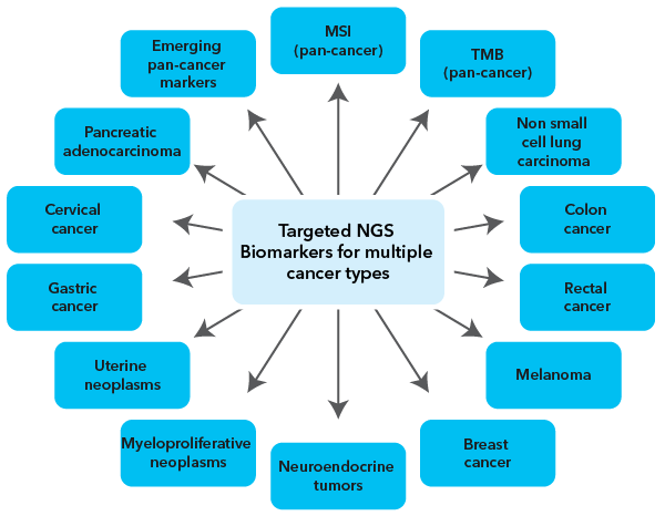
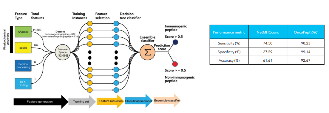
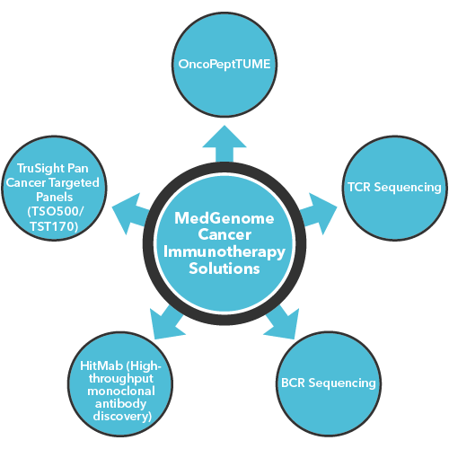
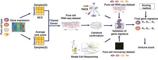
MedGenome offers advanced NGS solutions to accelerate lung cancer research, specializing in streamlined workflows and single-cell sequencing as a certified 10x Genomics provider. Our services include expert consultation, efficient sample processing, rapid data delivery, and customized visualizations. We leverage Illumina TruSight Oncology 500 (TSO500) for comprehensive genomic profiling, identifying key biomarkers like microsatellite instability (MSI) and TMB, as well as detecting fusions, indels, SNVs, and amplifications. Our powerful bioinformatics platform transforms raw sequencing data into high-quality, actionable reports, driving faster breakthroughs in understanding lung cancer and guiding the development of precision therapies. Additionally, our proprietary algorithm, OncoPeptTUME™ maps the tumor microenvironment using RNA-seq data, offering insights into tumor heterogeneity and immune interactions, customizable for cancer immunotherapy research.

Please reach out to our expert scientific team at research@medgenome.com for any questions or further information.

To know more about our advanced genomics solutions and services please click on the following links: Spatial transcriptomics, Single cell sequencing, RNA sequencing, Immune profiling, Cancer panels, Whole genome and whole exome sequencing and Epigenetic profiling


References

    • Liu H, Pan W, Tang C, Tang Y, Wu H, et al. The methods and advances of adaptive immune receptors repertoire sequencing. Theranostics. 11, 8945-8963 (2021).
    • Padinharayil H, Alappat RR, Joy LM, Anilkumar KV, Wilson CM, et al. Advances in the Lung Cancer Immunotherapy Approaches. Vaccines (Basel). 10, 1963 (2022).
    • Lahiri A, Maji A, Potdar PD, Singh N, Parikh P, et al. Lung cancer immunotherapy: progress, pitfalls, and promises. Mol Cancer. 22, 40 (2023).
    • Yan Y, Shen S, Li J, Su L, Wang B, et al. Cross-omics strategies and personalised options for lung cancer immunotherapy. Front Immunol. 15, 1471409 (2024).
    • Mamdani H, Matosevic S, Khalid AB, Durm G, Jalal SI. Immunotherapy in Lung Cancer: Current Landscape and Future Directions. Front Immunol. 13, 823618 (2022).
    • Altorki NK, Bhinder B, Borczuk AC, Elemento O, Mittal V, McGraw TE. A signature of enhanced proliferation associated with response and survival to anti-PD-L1 therapy in early-stage non-small cell lung cancer. Cell Rep Med. 5, 101438 (2024).
    • Xiao N, Liu H, Zhang C, Chen H, Li Y, et al. Applications of single-cell analysis in immunotherapy for lung cancer: Current progress, new challenges and expectations. J Adv Res. S2090-1232(24)00462-4 (2024).
    • Zhou X, Wang N, Zhang Y, Yu H, Wu Q. KAT2B is an immune infiltration-associated biomarker predicting prognosis and response to immunotherapy in non-small cell lung cancer. Invest New Drugs. 40, 43-57 (2022).


#Lung cancer, #Lung adenocarcinoma, #Non-small cell lung cancer (NSCLC), #Small cell lung cancer (SCLC), #EGFR, #ALK, #Immunotherapy, #Immune checkpoint inhibitors, #PD-L1, #Biomarkers, #tumor microenvironment, #Circulating tumor DNA, #Single cell RNA sequencing, #Tumor mutation burden, #TSO500
 

Immune repertoire diversity: Key to understanding disease processes and creating novel therapies

The adaptive immune system’s ability to recognize and neutralize a vast array of pathogens relies on the diversity of its lymphocyte repertoire. T and B lymphocytes, with their unique T cell receptors (TCRs) and B cell receptors (BCRs), are crucial for immunological memory and effective immune responses. This diversity allows the immune system to identify and combat a broad range of pathogens. Studying immune repertoire diversity offers valuable insights into disease mechanisms, therapeutic strategies, and vaccine development.

By Dr. Lavanya Balakrishnan and Vinay C. G., MedGenome Scientific Affairs

The adaptive immune system’s ability to recognize and neutralize a vast array of pathogens relies on the diversity of its lymphocyte repertoire. T and B lymphocytes, with their unique T cell receptors (TCRs) and B cell receptors (BCRs), are crucial for immunological memory and effective immune responses. This diversity allows the immune system to identify and combat a broad range of pathogens. Studying immune repertoire diversity offers valuable insights into disease mechanisms, therapeutic strategies, and vaccine development.

How TCRs and BCRs differ and contribute to immune repertoire diversity

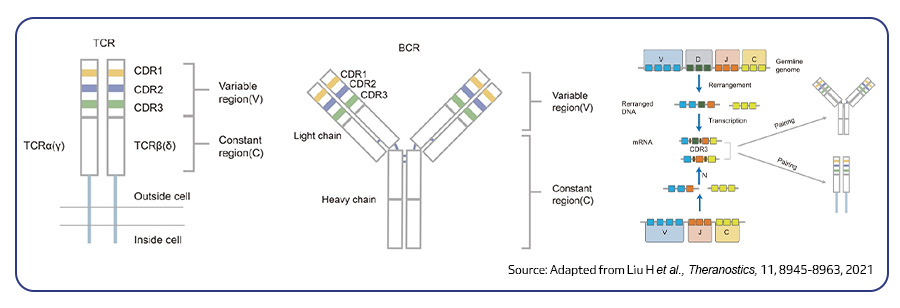
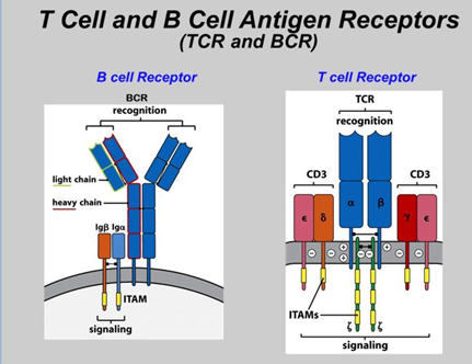
TCRs recognize antigenic peptides presented by MHC molecules, while BCRs and antibodies bind directly to antigen surfaces. TCRs consist of either α and β chains or γ and δ chains, with most T cells expressing αβ TCRs and a smaller percentage expressing γδ TCRs involved in innate immunity. BCRs are membrane-bound and consist of heavy and light chains, while immunoglobulins (Ig) are secreted by B/plasma cells1,2.

These immune receptors arise from somatic V(D)J recombination, generating over 1018 potential diversities in T cells and 1013 in B cells. Each receptor is composed of variable (V), diversity (D), joining (J), and constant (C) gene segments, which determine lymphocyte specificity. TCRs and BCRs include three complementarity-determining regions (CDR1, CDR2, CDR3), with CDR3 being crucial for antigen binding. Recombination involves joining D and J segments first, then V, with exonucleases removing and random nucleotides being added, enhancing junctional diversity. The D segment is present only in TCR β, TCR γ, and BCR heavy chains, while other chains only involve V and J segments. B cell receptors may also undergo somatic hypermutation and class-switch recombination to enhance antibody affinity and diversify the immune repertoire1,2.

Structure of TCR and BCR
Figure 1: Structure of (A)TCR, (B) BCR and (C) The mechanism of lymphocyte receptor diversity

Leveraging immune repertoire sequencing for innovative therapeutic strategies

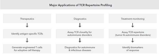
Immune repertoire profiling, through TCR and BCR sequencing, provides a comprehensive view of the immune system. By analyzing clonal diversity and dynamics, researchers gain insights into immune responses, disease mechanisms, and therapeutic targets. This approach is transforming our understanding of immunity and driving advancements in immunotherapy, immuno-oncology, autoimmune, and infectious diseases. Several studies have validated the utility of immune repertoire sequencing for investigating fundamental questions in healthcare and medical science. Summarized below are a few studies that highlight the benefits of bulk TCR/BCR sequencing in advancing our understanding of immune responses.

Overlapping T cell signatures in blood and tumor correlate with PD-1 efficacy

T cell receptor repertoire predicts response to PD-1 immunotherapy. Given the increasing use of PD-1 blockade, identifying predictive biomarkers is crucial. TCR repertoire analysis of gastrointestinal cancer cases who have undergone treatment with anti-PD1 antibody (nivolumab) revealed a correlation between treatment response and the overlap of T cell clones found in blood and tumor tissue. Individuals with a higher frequency of shared T cell clones in their blood before treatment experienced better clinical outcomes. These findings suggest that TCR repertoire analysis can potentially serve as a predictive biomarker to guide stratification of individuals for PD-1 therapy3.

Characterization of TCR and BCR repertoires in hepatocellular carcinoma

In hepatocellular carcinoma (HCC), tumor-infiltrating T and B cells play a crucial role in anti-tumor immunity. Analyzing immune repertoire (IR) features of tumor and non-tumor tissues from 64 HCC cases revealed high IR heterogeneity, with non-tumor tissues showing higher BCR diversity and somatic hypermutation, while tumor tissues had comparable or higher TCR diversity and lower immune infiltration. Notably, higher IR evenness in tumors and lower TCR richness in non-tumor tissues correlated with better patient survival. These findings suggest that IR features could serve as biomarkers for HCC diagnosis and treatment, aiding future immunotherapy strategies4.

COVID-19 severity linked to T and B Cell receptor profiles

By comprehensively profiling the immune responses of individuals infected with SARS-CoV-2, researchers constructed a vast repository of B and T cell receptor sequences. Analysis of this data revealed distinct patterns in antibody and T cell responses correlated with disease severity and progression. Specific B cell clusters associated with virus-neutralizing antibodies were identified, along with diverse T cell responses involved in early immune activation, antiviral responses, and regulatory functions. These findings provide a foundation for developing effective vaccines and immunotherapies against SARS-CoV-25.

Decode immune repertoires with MedGenome’s immune profiling solutions

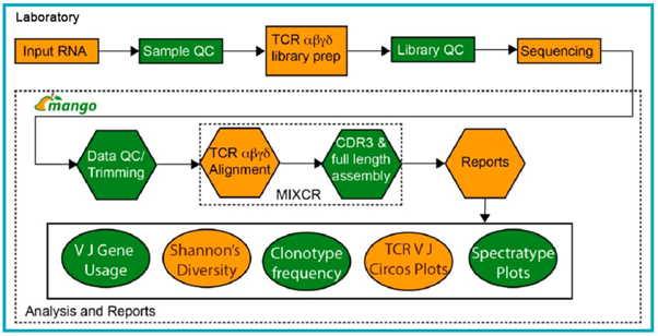
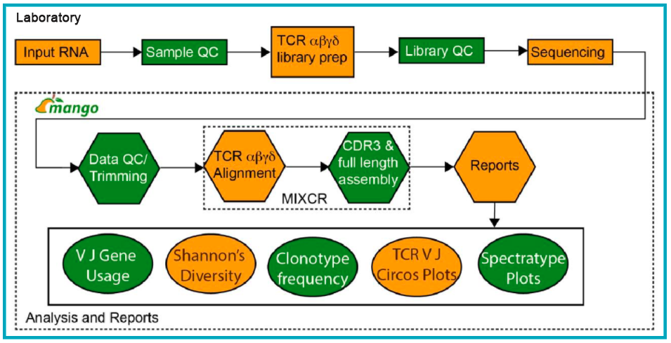
MedGenome delivers advanced immune profiling solutions to unravel the intricacies of TCR and BCR repertoires. Our comprehensive platform integrates high-throughput technology with expert bioinformatics, providing precise and actionable insights. From sample preparation to in-depth repertoire characterization, including the detection of rare clonotypes, our seamless workflow empowers researchers to accelerate discoveries. Our expert team offers tailored support and generates publication-ready results. Utilizing Takara’s Immuneprofiler and VDJ tools, our analysis pipeline delivers comprehensive reports encompassing full-length clonotype sequences, clonal frequencies, diversity metrics, V and J gene usage, and phylogenetic analysis of targeted clonotypes.

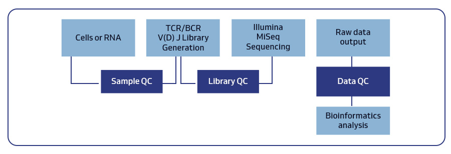
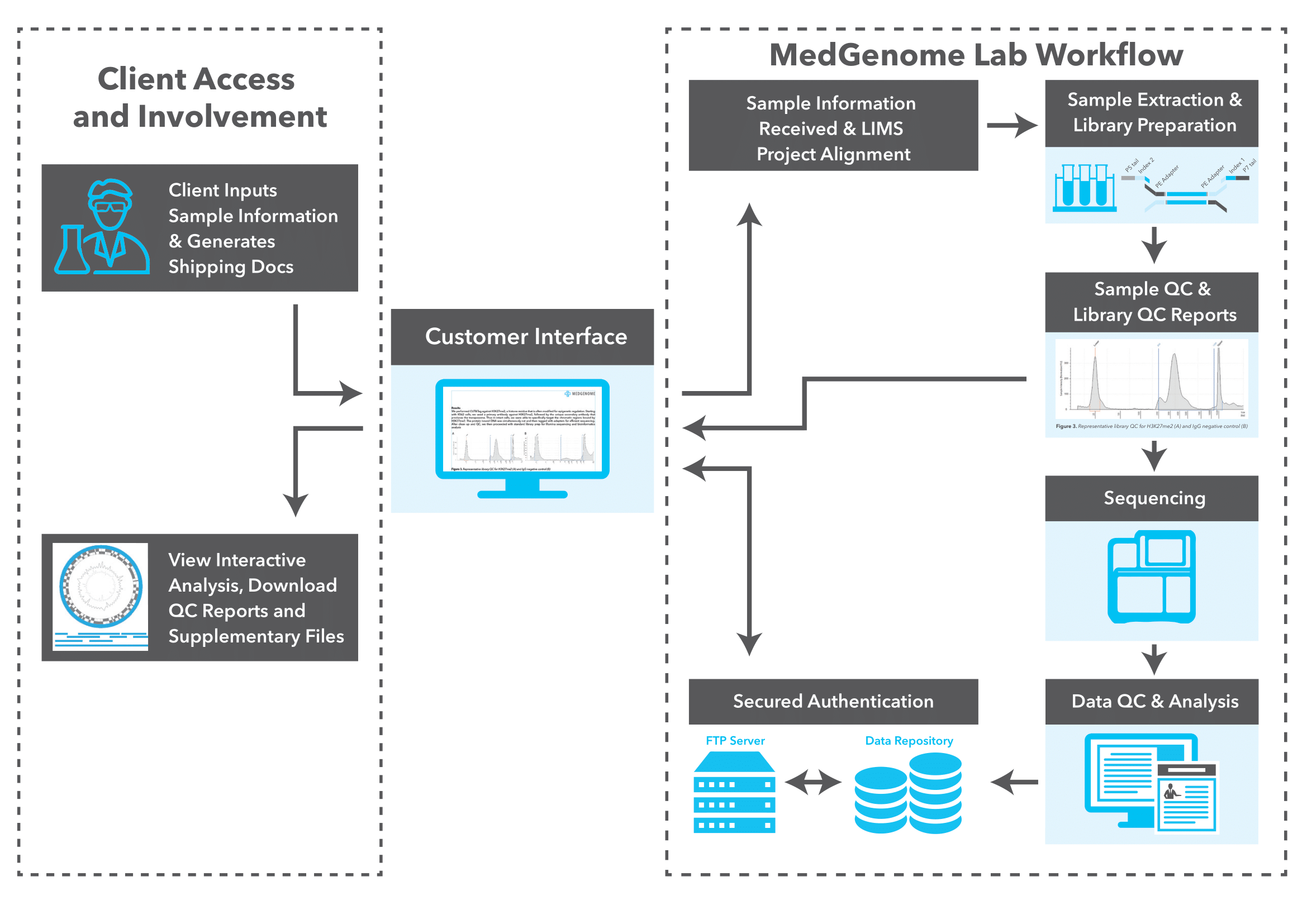
Workflow for bulk TCR and BCR profiling
Figure 2: End-to-end service workflow for bulk TCR and BCR profiling offered by MedGenome


Table 1. Details of sample types, library generation methods and analysis offerings for bulk TCR and BCR profiling
TCR BCR
Sample type Cells, tissue, blood and purified RNA Cells and purified RNA
Sample requirement >10ng Total RNA; >50 cells 10ng-3μg RNA; 1000-10,000 cells
Library generation method Takara SMARTer TCR alpha/beta Profiling Kit (Human, Mouse and custom species) Takara SMARTer BCR IgG/M Profiling Kit (Human and Mouse)
Analysis offerings Standard Analysis

  • FASTQ
  • MiXCR output file – Clonotype frequency, gene and amino acid sequences
  • V J gene usage
  • Shannon’s diversity
  • V J Circos plot
  • Spectratype plot
  • Scatter plot
  • Custom figures available upon request
Standard Analysis

  • FASTQ
  • MiXCR output file – Clonotype frequency, gene and amino acid Sequences
  • V J gene usage, Circos plots and Spectratype plot
  • Custom figures available upon request

MedGenome’s comprehensive and precise immune repertoire analyses enables the identification of critical biomarkers and therapeutic targets, empowering researchers to develop innovative treatments for cancer, infectious diseases, and autoimmune disorders.

Optimize your immune research with MedGenome’s advanced repertoire profiling. Contact us to see how our immune profiling and antibody discovery services can accelerate your R&D efforts.


References

    • Liu H, Pan W, Tang C, Tang Y, Wu H, et al. The methods and advances of adaptive immune receptors repertoire sequencing. Theranostics. 11, 8945-8963 (2021).
    • Katoh H, Komura D, Furuya G, Ishikawa S. Immune repertoire profiling for disease pathobiology. Pathol Int. 73, 1-11 (2023).
    • Aoki H, Ueha S, Nakamura Y, Shichino S, Nakajima H, et al. Greater extent of blood-tumor TCR repertoire overlap is associated with favorable clinical responses to PD-1 blockade. Cancer Sci. 112, 2993-3004 (2021).
    • Xie S, Yan R, Zheng A, Shi M, Tang L, et al. T cell receptor and B cell receptor exhibit unique signatures in tumor and adjacent non-tumor tissues of hepatocellular carcinoma. Front Immunol. 14, 1161417 (2023).
    • Schultheiß C, Paschold L, Simnica D, Mohme M, Willscher E, et al. Next-Generation Sequencing of T and B Cell Receptor Repertoires from COVID-19 Patients Showed Signatures Associated with Severity of Disease. Immunity. 53, 442-455.e4 (2020).


#Immune repertoire sequencing, #T-cell receptor, #B-cell receptor, #TCR, #BCR, #Adaptive immune system, #V(D)J recombination, #Somatic hypermutation, #Class switch recombination, #Immunotherapy, #COVID-19, #Biomarkers, #Immuno-oncology, #Autoimmune diseases, #Antibody discovery


Comprehensive cancer profiling with Illumina’s TSO 500 assay

Illumina’s TSO 500 empowers comprehensive genomic profiling (CGP), unlocking crucial tumor biomarkers to drive precision medicine. By focusing on clinically relevant genomic regions, CGP provides in-depth analysis, accurately detects low-frequency mutations, and comprehensively characterizes tumors. This facilitates tailored treatment approaches, guiding therapy selection based on specific molecular profiles. Moreover, CGP enables non-invasive disease monitoring through circulating tumor DNA (ctDNA) analysis.
By Dr. Lavanya Balakrishnan and Vinay C. G., MedGenome Scientific Affairs

Illumina’s TSO 500 empowers comprehensive genomic profiling (CGP), unlocking crucial tumor biomarkers to drive precision medicine. By focusing on clinically relevant genomic regions, CGP provides in-depth analysis, accurately detects low-frequency mutations, and comprehensively characterizes tumors. This facilitates tailored treatment approaches, guiding therapy selection based on specific molecular profiles. Moreover, CGP enables non-invasive disease monitoring through circulating tumor DNA (ctDNA) analysis.
TSO 500: Advanced NGS assay for in-depth pan-cancer genomic profiling

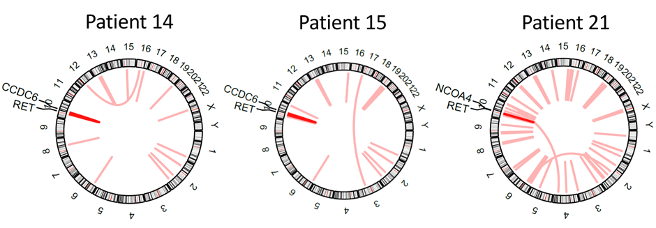
TSO 500 is a cutting-edge next-generation sequencing (NGS) assay designed for pan-cancer genomic profiling. TSO 500 targets the full coding regions of 523 genes known to be implicated in cancer and offers a comprehensive analysis of genetic alterations in solid tumors including single nucleotide variants (SNVs), insertions/deletions (InDels), copy number variations (CNVs), gene fusions and splice variants. Furthermore, TSO 500 also assesses microsatellite instability (MSI) and tumor mutational burden (TMB), biomarkers crucial for understanding the tumor’s behavior and potential response to immunotherapy1.
Variant types detected by TSO 500 solutions
Figure 1: Variant types detected by TSO 500 solutions

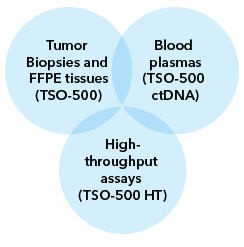
TSO 500 employs a hybridization-based capture approach using unique molecular identifiers (UMIs) and allows the analysis of both DNA and RNA from a single sample1. The TSO 500 portfolio offered by MedGenome includes two assays: TSO 500 for tissue-based profiling and TSO 500 ctDNA for liquid biopsy analysis.

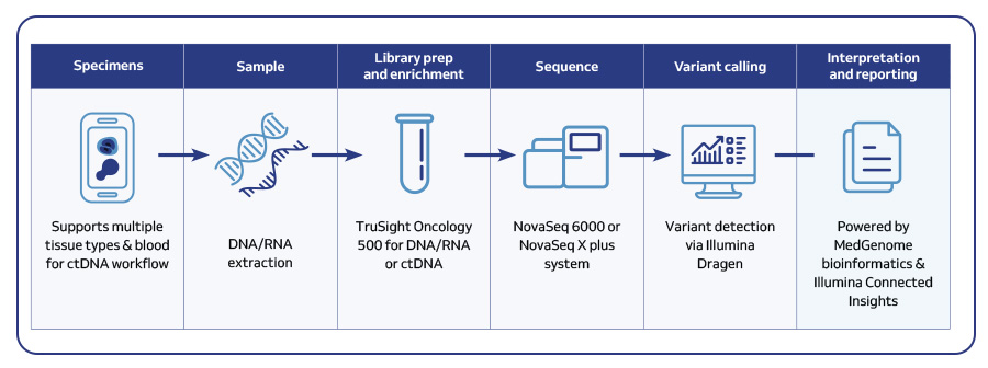
MedGenome’s workflow for comprehensive tumor profiling with TSO 500
Figure 2: MedGenome’s workflow for comprehensive tumor profiling with TSO 500


Table. 1. Features of TSO 500 portfolio offered by MedGenome
TSO 500 TSO 500 ctDNA
Sample type Tissue biopsies and FFPE Blood
Sample input and amount DNA and RNA; 50 ng purified DNA and RNA Cell free DNA; 20–30 ng purified ctDNA
Genes covered 523 genes for DNA variants; 55 genes for RNA fusions and splice variants 523 genes for DNA variants; 23 genes for DNA fusions
Variants called and Genomic signatures
  • SNVs
  • InDels
  • CNVs
  • Fusions (RNA)
  • Splice variants
  • TMB
  • MSI
  • HRD (DNA)
  • SNVs
  • InDels
  • CNVs
  • TMB
  • MSI
Panel size 1.94 Mb DNA; 358 kb RNA 1.94 Mb DNA
Sequencing platform and read depth Novaseq 6000 or Novaseq X plus
DNA: >80 M PE Reads; 100 bp PE
RNA: >40 M PE Reads; 100 bp PE
Novaseq 6000 or Novaseq X plus
>400 M PE Reads; 150 bp PE
Approximate turnaround time 5-7 days 5-7 days
Sample batching TSO 500 – 8 samples/run
TSO 500 HRD – 8-16 samples/run
8–48 samples/run
Analytical specificity 99.9998% 99.9994% (SNVs)
Analytical sensitivity >96% >95%


Predictive biomarker identification: TMB and MSI with TSO 500

The TSO 500 assay is a comprehensive solution for identifying patients likely to benefit from immunotherapy. By accurately quantifying TMB and determining MSI status, two key predictive biomarkers, the assay allows to make informed treatment decisions. Leveraging error-corrected sequencing and robust bioinformatics, the assay enables precise measurement of TMB, including both synonymous and nonsynonymous mutations, and reliable assessment of MSI. Both TMB and MSI values generated by this assay demonstrated high concordance with those obtained from whole exome sequencing and PCR-based assays, respectively.

From data to insights: Illumina Connected Insights and Dragen

Illumina Connected Insights, powered by the Dragen bioinformatics platform, transforms complex genomic data into actionable insights. By integrating the TSO 500 assay, this powerful combination delivers comprehensive tumor profiling, enabling precise patient selection and optimized treatment strategies. Dragen rapidly processes vast amounts of sequencing data, providing accurate variant calls, while Illumina Connected Insights offers a user-friendly interface for interpretation, clinical decision support, and seamless workflow integration.

Effectiveness of TSO 500 in cancer immunotherapy

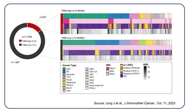
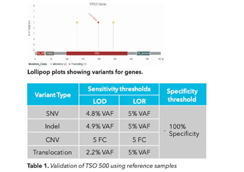
The TSO 500 assay has proven highly effective in immunotherapy by identifying biomarkers such as TMB and MSI that predict response to immune checkpoint inhibitors. Several studies have shown how the assay has helped identify actionable mutations in various types of cancer, leading to successful targeted therapies. Validation of TSO 500 on 170 clinical samples across different cancers demonstrated precision and accuracy of over 99%, with sensitivity and specificity of at least 99% for all variant types2. Using this assay, higher response rates to anti-PD-(L)1 therapy in TMB-high cases were observed, particularly in gastric, gallbladder, head and neck cancers, and melanoma3. It also highlighted significant genetic alterations, such as BRAF mutations in peritoneal metastases from colorectal cancer4 and heterogeneity in intrahepatic cholangiocarcinoma5. Additionally, TSO 500’s comprehensive profiling of early-stage NSCLC6 and endometrial serous carcinoma7 revealed actionable mutations and potential markers for prognosis and treatment stratification. These findings underscore TSO 500’s role in enhancing diagnostic accuracy and guiding therapeutic decisions across different cancer types.

Clinical and molecular characteristics of TMB-High and TMB-Low patient
Figure 3: Clinical and molecular characteristics of TMB-High and TMB-Low patient cohorts along with PD-L1 expression profiles.


How MedGenome’s comprehensive genomic profiling enhances targeted therapies

MedGenome offers end-to-end customized genomic profiling solutions to accelerate cancer research. Our expertise in handling diverse sample types, including tumor biopsies, FFPE tissues, and liquid biopsies, combined with advanced bioinformatics capabilities, ensures rapid and accurate analysis. With rapid turnaround times and scalable operations, MedGenome empowers researchers to unlock the potential of precision oncology. We analyze TSO 500 data using Dragen with Illumina Connected Insights, delivering the highest accuracy in variant calls with the fastest analysis time. Our variant summary report includes results from Dragen and Illumina Connected Insights, with advanced analysis reports featuring rich visualizations of mutations, fusions, copy number alterations, and immuno-oncology biomarkers such as TMB and MSI.

Contact us now to learn how our TSO Targeted sequencing can drive your research forward.


References

    • https://sapac.illumina.com/products/by-brand/trusight-oncology/tso-500-portfolio.html
    • Froyen G, Geerdens E, Berden S, Cruys B, Maes B. Diagnostic Validation of a Comprehensive Targeted Panel for Broad Mutational and Biomarker Analysis in Solid Tumors. Cancers (Basel), 14, 2457 (2022).
    • Jung J, Heo YJ, Park S. High tumor mutational burden predicts favorable response to anti-PD-(L)1 therapy in patients with solid tumor: a real-world pan-tumor analysis. J Immunother Cancer., 11, e006454 (2023).
    • Heuvelings DJI, Wintjens AGWE, Moonen L, Engelen SME, de Hingh IHJT, et al. Predictive Genetic Biomarkers for the Development of Peritoneal Metastases in Colorectal Cancer. Int J Mol Sci., 24, 12830 (2023).
    • Kinzler MN, Schulze F, Jeroch J, Schmitt C, Ebner S, et al. Heterogeneity of small duct- and large duct-type intrahepatic cholangiocarcinoma. Histopathology, 84, 1061-1067 (2024).
    • Choi SJ, Lee JB, Kim JH, Hong MH, Cho BC, Lim SM. Analysis of tumor mutational burden and mutational landscape comparing whole-exome sequencing and comprehensive genomic profiling in patients with resectable early-stage non-small-cell lung cancer. Ther Adv Med Oncol., 16, 17588359241240657 (2024).
    • Aisagbonhi O, Ghlichloo I, Hong DS, Roma A, Fadare O, et al. Comprehensive next-generation sequencing identifies novel putative pathogenic or likely pathogenic germline variants in patients with concurrent tubo-ovarian and endometrial serous and endometrioid carcinomas or precursors. Gynecol Oncol., 187, 241-248 (2024).


#Tumor profiling, #Next-generation sequencing, #Targeted sequencing, #circulating tumor DNA, #ctDNA, #TSO 500, #TSO 500 ctDNA, #Tumor mutational burden (TMB), #Microsatellite instability (MSI), #Liquid biopsies, #Error corrected sequencing, #Illumina Connected Insights, #Dragen, #Precision oncology, #Targeted therapies


Single-Cell Sequencing: Applications, Methods, and Insights

Single-cell sequencing offers unprecedented resolution for analyzing the unique molecular signatures of individual cells. By deconstructing the cellular landscape, it reveals hidden cell populations, tracks how cells differentiate, and identifies disease biomarkers. By dissecting the genomic, transcriptomic, and epigenetic landscape of single cells, researchers can now gain a deeper understanding of complex biological processes, disease mechanisms, and how patients respond to treatments. This method is essential for both basic and clinical research for unlocking the mysteries of cellular complexity and dynamic biological processes.

By Dr. Lavanya Balakrishnan, Vinay C. G. and Michelle Balakrishnan Vierra, MedGenome Scientific Affairs

Single-cell sequencing offers unprecedented resolution for analyzing the unique molecular signatures of individual cells. By deconstructing the cellular landscape, it reveals hidden cell populations, tracks how cells differentiate, and identifies disease biomarkers. By dissecting the genomic, transcriptomic, and epigenetic landscape of single cells, researchers can now gain a deeper understanding of complex biological processes, disease mechanisms, and how patients respond to treatments. This method is essential for both basic and clinical research for unlocking the mysteries of cellular complexity and dynamic biological processes.

Single-cell sequencing: techniques and practical applications in cellular research

Single-cell sequencing utilizes a variety of techniques, with each assay tailored to target specific cellular information according to the investigator’s needs. Here’s an overview of key techniques and their applications:

Single-cell RNA sequencing (scRNA-seq)

scRNA-seq enables gene expression analysis at the single-cell level, revealing hidden cellular diversity. By profiling RNA molecules, it identifies new cell types and discovers disease-associated gene networks, facilitating biomarker discovery and treatment improvement. This technique accelerates drug development by enabling precise patient stratification and monitoring, leading the way to personalized medicine.

Case Example: Recent MedGenome-Supported Publication

A recent study by Pasqualina Colella et al. from Stanford University, published in Nature, employed scRNA-seq to reveal unexpected diversity in microglia-like cells within the brain. This finding shed light on how blood-derived cells can repopulate the central nervous system. Their work further demonstrates the therapeutic potential of stem cell transplants for progranulin-deficient neurodegenerative diseases1.

scRNA seq analysis of brain and bone
Figure 1: scRNA seq analysis of brain and bone marrow cells reveal the heterogeneity of microglial like cells. (A) Experimental design showing scRNA-seq analysis of different kinds of cells isolated from mice. (B) UMAP projection depicting the clustering of cells. (C) Differential gene expression of microglia signature genes depicted using dot plot.

Single-cell DNA sequencing (scDNA-seq)

scDNA-seq is highly useful in understanding the genetic makeup of individual cells, identify driver mutations and copy number variations to reveal tumor heterogeneity and cancer evolution. One interesting publication by Ng et al. (2024) employed scDNA-seq to investigate the mechanisms leading to amplicons in esophageal adenocarcinoma, a cancer fueled by frequent gene amplifications. This advanced approach provided deeper insights into how amplified regions contribute to tumor evolution over time, offering valuable understanding of cancer progression2.

Frequency of amplified genomic regions in esophageal adenocarcinoma
Figure 2: Frequency of amplified genomic regions in esophageal adenocarcinoma.

Single-cell TCR/BCR sequencing

Single-cell TCR/BCR sequencing technique is highly useful in understanding the immune system’s adaptive defenses by profiling T and B cell populations, uncovering rare immune cells crucial for fighting infections and tumors. The technique aids in analyzing unique variable regions within T and B cell receptors, enabling researchers to map antigen specificities and track immune cell development. Blanco-Heredia et al, Memorial Sloan Kettering Cancer Center, New York, used single-cell TCR sequencing to explore how the immune response interacts with tumor evolution in triple-negative breast cancer. The study found that as cancer progresses, the immune response weakens, marked by declining T cell diversity and the emergence of tumor escape mechanisms3.

UMAP analysis of peripheral blood TCR clonotypes
Figure 3: UMAP analysis of peripheral blood TCR clonotypes from a TNBC patient reveals the evolution of TCR repertoire.

Single-cell ATAC-seq (scATAC-seq)

scATAC-seq decodes gene regulation within individual cells, identifying unique regulatory patterns and shedding light on cellular differentiation mechanisms. A study by Terekhanova et al. (2023) in Nature highlighted the impact of chromatin accessibility on cancer. Analyzing over 1 million cells from 225 tumor samples across 11 cancer types, the researchers found that changes in DNA accessibility can initiate, progress, and metastasize cancer. They identified both common and cancer-specific gene regulation patterns, including key pathways like TP53, hypoxia, and TNF signaling. The study also emphasized the role of estrogen response and epithelial-mesenchymal transition in metastasis, linking DNA accessibility to gene expression and cancer dynamics4.

top cancer-cell-associated
Figure 4: The top cancer-cell-associated differentially accessible chromatin regions identified across 11 cancer types.

Single-cell multi-omics analysis

Single-cell multi-omics analysis integrates diverse data types beyond gene expression (scRNA-seq), including cell surface proteins, antigen receptors (TCR/BCR), and chromatin accessibility (scATAC-seq). CITE-seq further empowers this approach by simultaneously analyzing a vast number of cell surface proteins alongside gene expression in a single cell, offering a deeper understanding of cellular interactions and regulation. For example, a study using scRNA-seq and scATAC-seq on pediatric KMT2A-rearranged leukemia revealed key insights for targeted as well as immunotherapies5.

Heatmap
Figure 5: Heatmap of (A) differentially expressed genes and (B) enriched transcription factors identified from pediatric KMT2A-rearranged leukemia.

A seamless workflow for new discoveries

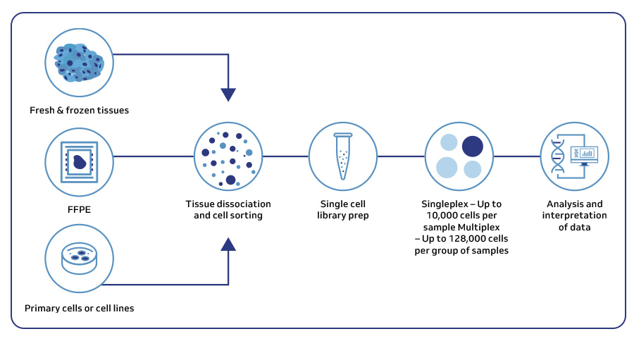
As a trusted 10x Genomics certified provider, MedGenome, offers end-to-end solutions tailored to your specific single-cell research needs. From sample preparation to data analysis, our expert team is there to support your project.

General workflow for single-cell sequencing analysis
Figure 6: A general workflow for single-cell sequencing analysis using 10x Genomics offered by MedGenome.

Extracting meaning from the data: Bioinformatics tools

At MedGenome, we have the expertise and the extensive computational infrastructure to provide a seamless experience from sample through insightful data analysis.

MedGenome offers a range of bioinformatics analysis options to meet your specific research needs:

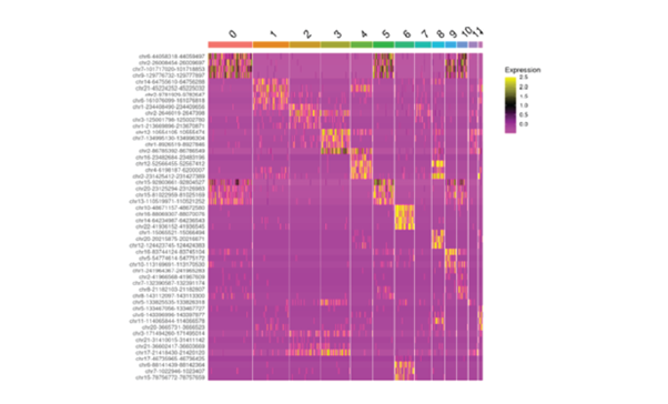
  • Standard analysis: We provide raw sequencing data (FASTQ files) and insights from Cell Ranger, including quality control metrics, gene expression levels, and heatmap visualizations for initial exploration using Loupe Browser software.
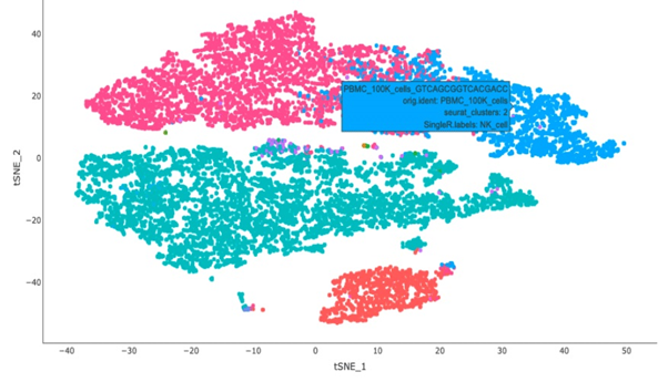
  • Advanced analysis: MedGenome’s advanced analysis capabilities include all standard analysis deliverables along with advanced QC via Seurat and enhanced filtering of low-quality cells, contamination, and multiplets. The analysis also encompasses dimensionality reduction and clustering analysis on filtered data, interactive t-SNE plots with cluster information, and general cell type annotation. Additionally, MedGenome offers differential gene expression analysis for clusters and annotated cell types, pathway enrichment analysis, and group comparison.
  • Specialized analysis: Tailor the analysis further with custom cell type annotations based on your unique markers. Uncover functional differences between cell types through differential gene expression analysis and map their interactions via cell-to-cell interactome analysis, providing a more comprehensive understanding of cellular behavior.
UMAP plot
Figure 7: (A) UMAP plot of gene expression data annotated by cell type. (B) Bubble plot of marker genes by cell type/cluster. (C) Clonotype phylogenetic tree from single cell TCR/BCR data analysis.

MedGenome, your single cell sequencing partner

MedGenome, a certified 10x Genomics partner, offers flexible and scalable single-cell sequencing solutions. We specialize in analyzing diverse samples—from fresh to cryopreserved and fixed specimens—using a range of advanced techniques. Our meticulous approach includes gentle tissue dissociation, viability checks, and precise cell sorting via FACS. Beyond processing, our expert bioinformatics team integrates samples, performs tailored analysis, and provides comprehensive, publication-ready reports. Researchers benefit from detailed metrics, plots, and raw data access for further exploration.

Unlock the full potential of your research with MedGenome’s cutting-edge single-cell sequencing solutions. Contact us today to learn how we can accelerate your scientific discoveries and drive groundbreaking insights in biology and medicine.


References

    • Colella P, Sayana R, Suarez-Nieto MV, Sarno J, Nyame K, et al. CNS-wide repopulation by hematopoietic-derived microglia-like cells corrects progranulin deficiency in mice. Nat Commun 15, 5654 (2024).
    • Ng AWT, McClurg DP, Wesley B, Zamani SA, Black E, et al. Disentangling oncogenic amplicons in esophageal adenocarcinoma. Nat Commun, 15, 4074 (2024).
    • Blanco-Heredia, J., Souza, C.A., Trincado, J.L. et al. Converging and evolving immuno-genomic routes toward immune escape in breast cancer. Nat Commun 15, 1302 (2024).
    • Terekhanova, N.V., Karpova, A., Liang, WW. et al. Epigenetic regulation during cancer transitions across 11 tumour types. Nature 623, 432–441 (2023).
    • Chen C, Yu W, Alikarami F, Qiu Q, Chen CH, Flournoy J, et al. Single-cell multiomics reveals increased plasticity, resistant populations, and stem-cell-like blasts in KMT2A-rearranged leukemia. Blood 139(14), 2198-2211 (2022).


#Single cell sequencing, #Single cell RNA sequencing, #Single cell DNA sequencing, #Single cell ATAC-seq, #Single cell TCR/BCR sequencing, #CITE-seq, #Single cell multiomics, #10x Genomics, #Single cell biomarker discovery, #Single cell clustering analysis, #Dimensionality reduction, #Personalized immunotherapy, #Single-cell tumor heterogeneity


Complementing strides made with short-reads with PacBio long-read sequencing

Short-read sequencing technologies have, without a doubt, revolutionized genomics. The ability to look at genetic variants at the base pair level and compare gene expression levels between normal and other conditions has made it possible to diagnose more diseases, develop more robust crops, and protect the biodiversity here on Earth.

By Michelle Vierra Balakrishnan

Short-read sequencing technologies have, without a doubt, revolutionized genomics. The ability to look at genetic variants at the base pair level and compare gene expression levels between normal and other conditions has made it possible to diagnose more diseases, develop more robust crops, and protect the biodiversity here on Earth.

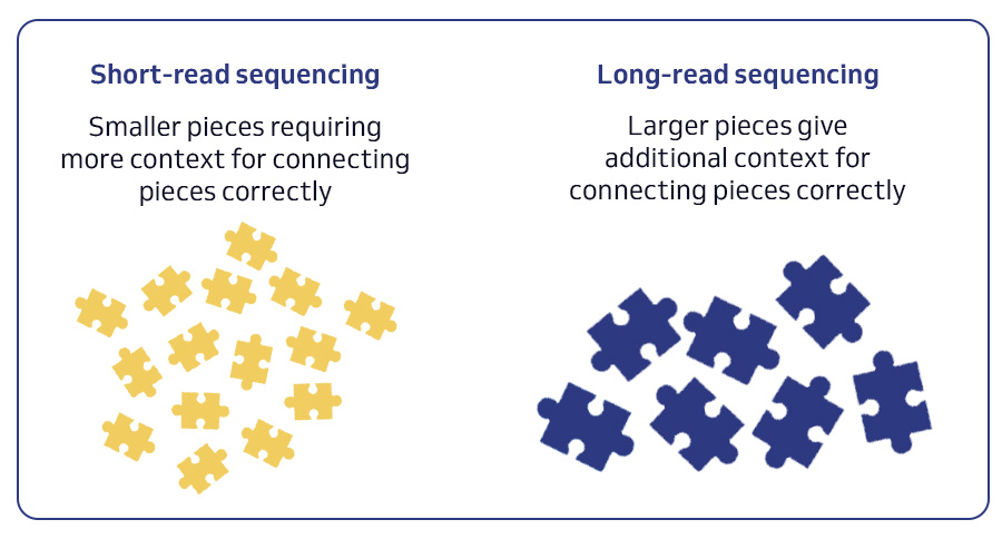
However, short reads are inherently limited and often fall short in fully characterizing complex genomes and transcriptomes. Short reads struggle to resolve repetitive regions, assemble complex structural variants, and fully capture isoform diversity. This is where the power of PacBio long-read sequencing comes into play.

PacBio HiFi sequencing delivers highly accurate long reads, enabling us to more fully profile not only the bases involved, but the context in which genetic variation exists. The ability to sequence every base in a genome or transcriptome opens doors previously blocked by extreme GC content, repeat rich regions, and long stretches of homozygosity. And as scientists dedicated to making the world a healthier place, MedGenome now offers researchers an unparalleled ability to delve deeper into the intricacies of genomes and transcriptomes with both short-read and long-read sequencing solutions.

Short and long read sequencing

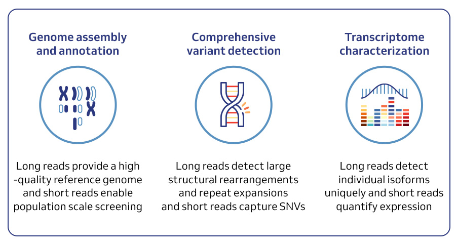
A comprehensive suite of PacBio solutions

  1. De novo genome assembly and annotation solution:
    • Generating high-quality, contiguous genome assemblies for any organism, regardless of complexity.
    • Identifying and annotating genes, isoforms, and regulatory elements with greater accuracy than ever before.
    • Understanding the complete genomic blueprint of an organism, crucial for evolutionary studies, conservation efforts, and agricultural advancement.
  2. Comprehensive human variant detection solution:
    • Detecting all types of genetic variation, including SNVs, Indels, SVs, and variants in complex regions, with high confidence.
    • Providing haplotype-resolved variant information, crucial for understanding inheritance patterns and disease mechanisms.
    • Enabling more accurate diagnosis, personalized treatment strategies, and a deeper understanding of human health and disease.
  3. Full-Length RNA sequencing solution:
    • Capturing the complete length of RNA transcripts, providing a comprehensive view of isoform diversity.
    • Identifying novel transcripts and fusion genes, crucial for understanding disease mechanisms and identifying potential drug targets.
    • Offering both bulk and single-cell full-length RNA sequencing, enabling researchers to dissect cellular heterogeneity and unravel the complexities of gene expression at single-cell resolution.

Combined capabilities – your omics superpower

The future of genomics lies in leveraging the strengths of both short-read and long-read technologies. This hybrid approach, combining the comprehensiveness of long reads with the affordability and counting ability of short reads, will undoubtedly accelerate discoveries and advance our understanding of complex biological systems.

Combined capabilities

From strategy to publication: crafting omic insights with your research in mind

At MedGenome, we believe in the power of comprehensive genomic exploration. That’s why we offer both short-read and PacBio long-read sequencing services, allowing you to choose the best strategy for your specific needs or combine both for unparalleled insights. Whether you’re assembling complex genomes, characterizing cryptic genetic variation, or unraveling the complexities of the transcriptome, our expert team is here to guide you every step of the way. Contact us today to discuss how our comprehensive suite of sequencing solutions can empower your next breakthrough discovery.


#De novo genome assembly and annotation, #human variant detection, #Full-Length RNA sequencing, #long-read sequencing, #pacbio


Navigating Spatial Data Analysis for Publishable Research

Spatial analysis has become an indispensable tool in unraveling the complexities of biological systems. From understanding gene function to dissecting tissue microenvironments, spatial data holds the key to unlocking valuable insights into biological processes. However, harnessing the full potential of spatial data requires not only sophisticated analytical techniques but also considerable computational resources and expertise.

By Michelle Vierra Balakrishnan

Spatial analysis has become an indispensable tool in unraveling the complexities of biological systems. From understanding gene function to dissecting tissue microenvironments, spatial data holds the key to unlocking valuable insights into biological processes. However, harnessing the full potential of spatial data requires not only sophisticated analytical techniques but also considerable computational resources and expertise.

Standard spatial analysis

At the heart of spatial analysis lies the extraction of meaningful information from spatially resolved transcriptomic data. However, the output of a spatial experimental run is a FASTQ file from a sequencer that has to be further analyzed for useful information. Space Ranger, the go-to solution for standard spatial analysis of 10x Genomics spatial data, provides researchers with essential statistics and visualizations to assess data quality and explore gene expression patterns.

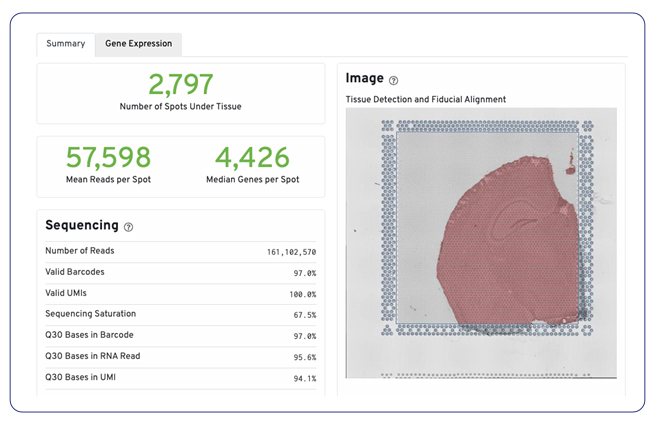
Space Ranger’s standard analysis pipeline generates key metrics such as the number of spots, genes per spot, and gene expression overlaid on slide images.

Example summary report from Space Ranger
Example summary report from Space Ranger.
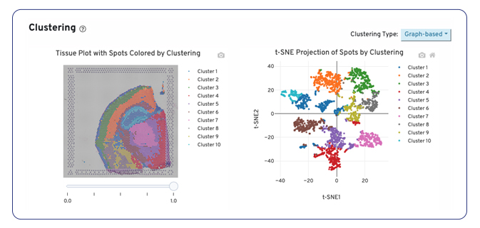
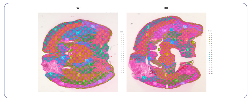
Example tissue plot and t-SNE projection of spots by clustering
Example tissue plot and t-SNE projection of spots by clustering.

While Space Ranger is a free tool, it does require expertise for installation and execution as well as significant computational resources to perform the analysis. For researchers who want to save the time and computational resources required, MedGenome offers this standard analysis as a nominal add on to your spatial project.

Advanced spatial analysis: getting multidimensional insights and publication-ready images for your tissue sections

To truly get valuable insights from spatial data, you need additional QC and biological context. MedGenome’s advanced spatial analysis solutions go beyond standard readouts, incorporating rigorous filtering criteria to ensure your data is high-quality and reliable. We first identify and filter out low-quality spots and contaminants such as mitochondrial and ribosomal RNA, providing a clean dataset.

Violin plots showing metrics used for filtering
Violin plots showing metrics used for filtering.

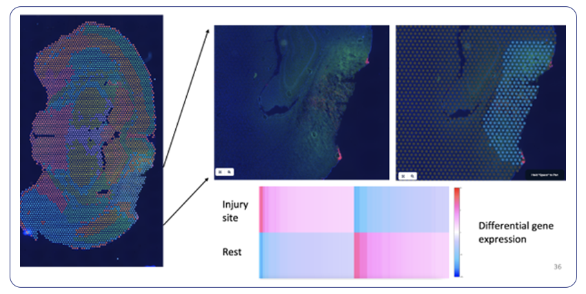
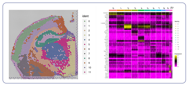
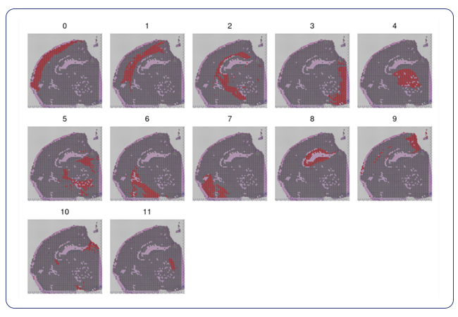
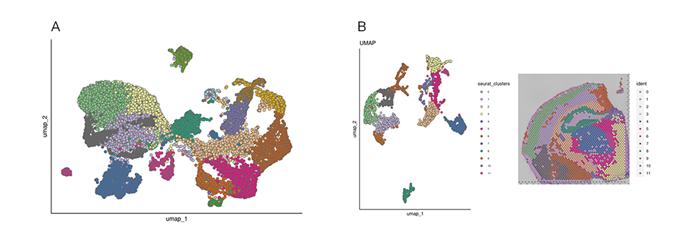
Following spot filtering, we use principal component analysis (PCA) and reclustering to provide a more accurate representation of the spatial transcriptome. We then provide visualizations as spatial plots by cluster to look at spatial localization of the spots. These plots, along with a heatmap and table of differential gene expression, furnish our clients with deeper insights into spatial gene expression patterns, paving the way for novel biological discoveries.

Spatial dimensional plot with filtered spots and heatmap of gene expression across clusters
Spatial dimensional plot with filtered spots and heatmap of gene expression across clusters.
Spatial localization of individual clusters
Spatial localization of individual clusters.

Specialized analysis: cell type annotation that fulfills your specific custom needs and interactome data for unique research questions

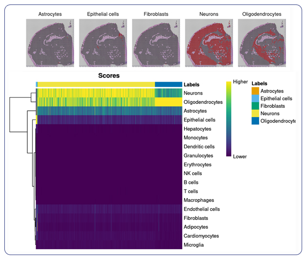
For researchers seeking specialized insights, MedGenome offers tailored analysis solutions designed to address specific research questions. Cell type annotation, a cornerstone of understanding the basis of tissue organization, is seamlessly integrated into MedGenome’s repertoire of analysis solutions. Leveraging public datasets, we can annotate the cell types in your data based on the tissue you are exploring. With this analysis, we provide spatial plots by cell type and provide additional plots for the confidence of cell annotation assignments.

Spatial localization by cell type and plot of confidence of assigned cell type annotation
Spatial localization by cell type and plot of confidence of assigned cell type annotation.

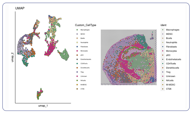
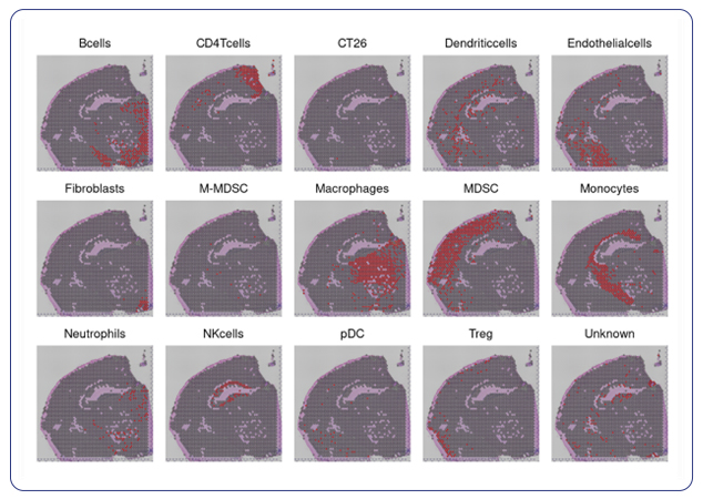
For researchers that have specific markers in mind for their cell types, we can take client-provided markers and annotate the specific cell types you’re interested in within your spatial data. With custom marker cell annotation we provide spatial plots by cell type and a table of differential gene expression.

UMAP and spatial dimensional plot with custom cell types
UMAP and spatial dimensional plot with custom cell types.
Spatial localization by cell type done with custom markers
Spatial localization by cell type done with custom markers.

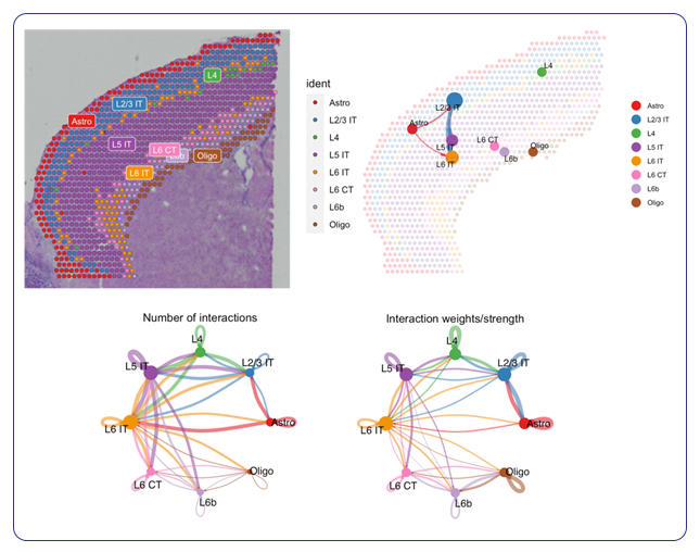
In addition to cell type annotation, MedGenome’s specialized analysis extends to pathway analysis and visualization of cell-to-cell interactions. Cell-to-cell communication models the probability of cell-to-cell communication by integrating gene expression with prior knowledge of the interactions between signaling ligands, receptors and their cofactors.

As part of the analysis, we provide a visualization of the interactome plotted on a spatial image showing not only the number of interactions between cell types, but also the weighted strength of those interactions. By elucidating molecular pathways and mapping intercellular communication networks, MedGenome equips researchers with a comprehensive understanding of tissue biology at the spatial level.

Spatial plot of pathways
Spatial plot of pathways, the interactions predicted across those spatial regions, and visualizations of the number of cell-to-cell interactions and the weights/strengths of those interactions.


From strategy to publication: crafting spatial insights with your research in mind

Spatial data analysis holds immense promise for advancing our understanding of complex biological systems. However, navigating the intricacies of spatial data analysis requires more than just off-the-shelf tools—it demands specialized expertise and tailored solutions. MedGenome’s comprehensive suite of spatial analysis services, ranging from standard analysis to specialized annotation and pathway analysis, offers researchers a streamlined pathway to unlocking the rich biological insights hidden within spatial data.

Ready to apply spatial transcriptomics to your research questions? Get in touch with a MedGenome expert to scope out your project.


#spatial analysis, #spatial transcriptomics, #cell type annotation, #t-SNE, #transcriptomic data


Melanoma & Skin Cancer Awareness Month: Genomics for developing effective therapies

Skin cancer is the most common type of cancer in the US. One in five Americans have the likelihood of developing skin cancer by the age of 70. Some common manifestations of skin cancer include basal cell carcinoma (BCC), squamous cell carcinoma (SCC) and melanoma. Research suggests over 5 million cases of skin cancer occur annually in the US alone. Non-melanoma skin cancers (basal cell carcinoma and squamous cell carcinoma) are the most common, followed by melanoma, the more aggressive form. Other types of skin cancer include Merkel cell cancer, cutaneous T-cell lymphoma, Kaposi sarcoma, skin adnexal tumors and sarcomas.

By MedGenome Scientific Affairs

Skin cancer is the most common type of cancer in the US. One in five Americans have the likelihood of developing skin cancer by the age of 70. Some common manifestations of skin cancer include basal cell carcinoma (BCC), squamous cell carcinoma (SCC) and melanoma. Research suggests over 5 million cases of skin cancer occur annually in the US alone. Non-melanoma skin cancers (BCC and SCC) are the most common, followed by melanoma, the more aggressive form. Other types of skin cancer include Merkel cell cancer, cutaneous T-cell lymphoma, Kaposi sarcoma, skin adnexal tumors and sarcomas.

Understanding skin cancer risks: Several factors are associated with risk of developing skin cancer, including chronic exposure to UV light, number and size of moles on the skin, fair skin, and light hair color. In addition, a familial history of skin cancer increases the likelihood of developing the disease due to genetic predisposition. Other risk factors include sunburn history, light complexion, eye color, and hair color, as well as certain skin conditions such as chronic inflammation, immune suppression, and environmental exposures like arsenic exposure and radiation1.

Genetic insights into melanoma’s aggressive nature

While non-melanoma skin cancer has a negligible impact on cancer mortality, melanoma accounts for most skin cancer-related deaths1.

Malignant melanoma arises from unchecked growth or damage to melanocytes. It is characterized by significant genetic heterogeneity, with tumors showcasing numerous genetic alterations and mutations. This complexity fuels the aggressiveness of melanoma, driving tumor progression, metastasis, and resistance to treatment. A study featured in Nature Cell Biology delved into the role of LAP1 protein in melanoma advancement. Elevated LAP1 levels in metastatic cells hint at its crucial role in cancer spread, as demonstrated in this research2, highlighting LAP1 as a pivotal regulator of melanoma’s aggressiveness. Targeting LAP1 emerges as a promising strategy to curb melanoma spread. Conducted by researchers from the Francis Crick Institute, Queen Mary University of London, and King’s College London, the study suggests classifying LAP1 as a potential prognostic marker in melanoma patients. Higher LAP1 levels at the perimeter of primary tumors correlate with increased melanoma aggressiveness and poorer outcomes.

Download Whitepaper button

Genetic factors and syndromic associations in skin cancer pathogenesis

Skin cancer occurrence is significantly influenced by hereditary genetic predisposition. For example:

  • Melanoma: Hereditary melanoma arises from mutations in two key genes, cyclin-dependent kinase inhibitor 2A (CDKN2A) and cyclin-dependent kinase 4 (CDK4), both major tumor suppressor genes. These mutations notably elevate melanoma risk, with CDKN2A mutations alone accounting for 35-40% of familial melanomas. Other implicated genes include BAP1, BRAF, CDK4, KIT, MITF, NF1, NRAS, PTEN, TERT, and TP53, affecting crucial pathways such as the phosphoinositide 3-kinase (PI3K)/AKT pathway and the RAS/RAF/MEK/ERK signaling cascade.
  • Basal cell carcinoma: Mutations in PTCH1 and PTCH2 genes underlie Basal Cell Nevus Syndrome, increasing the risk of BCC.
  • Squamous cell carcinoma: Certain syndromes like oculocutaneous albinism, epidermolysis bullosa, and Fanconi anemia are associated with higher susceptibility to SCC.

Current strategies for prevention and treatment of skin cancers:

Preventing skin cancer involves several key strategies aimed at reducing exposure to harmful UV radiation and minimizing other risk factors. These include using sunscreen, avoiding artificial sources of UV exposure like tanning beds and sunlamps, and regularly performing skin self-exams. By adopting these preventive measures, individuals can reduce their risk of developing skin cancer and promote overall skin health.

Early diagnosis is critical for skin cancer, as survival rates plummet with advanced disease stages. While surgery offers a cure for localized cases, advanced stages like metastatic melanoma have significantly worse outcomes, with 3-year overall survival ranging from a mere 4.7% to 26.4%. Fortunately, promising new avenues like immunotherapy and targeted therapies are providing renewed hope. BRAF/MEK inhibitors, anti-PD1 therapy, and combinations like nivolumab and ipilimumab have shown promising results in melanoma (Table 1). For advanced BCC, vismodegib and sonidegib target the Hedgehog pathway, while cemiplimab serves as a second-line therapy. Similarly, anti-PD1 agents like cemiplimab, pembrolizumab, and cosibelimab have yielded significant responses in locally advanced or metastatic SCC. PD-1/PD-L1 inhibitors and locoregional approaches are also under investigation for Merkel cell carcinoma3.

Additionally, Tumor mutation burden (TMB) analysis, facilitated by genomic techniques, is increasingly recognized as a pivotal determinant of treatment response, particularly in immunotherapy. High TMB correlates with heightened responsiveness to immunotherapeutic interventions, offering a promising avenue for personalized treatment strategies.

Table. 1. List of therapeutic agents approved by FDA for melanoma4
Sl. No. Therapeutic drug Mode of action
Targeted therapy
1 Vemurafenib BRAF inhibitor
2 Cobimetinib + Vemurafenib MEK inhibitor + BRAF inhibitor
3 Binimetinib + Encorafenib MEK inhibitor + BRAF Inhibitor
4 Dabrafenib + Trametinib BRAF inhibitor + MEK inhibitor
Immunotherapy
5 Ipilimumab Antibody against CTLA-4
6 Nivolumab Antibody against PD-1
7 Pembrolizumab Antibody against PD-1
8 Talimogene Laherparepvec (T-VEC) Oncolytic virus
9 Ipilimumab + Nivolumab Antibody against CTLA-4 + antibody against PD-1
10 Tebentafusptebn T-cell receptor-bispecific molecule that targets both glycoprotein 100 and CD3
11 Nivolumab + Relatlimab Antibody against PD-1 + antibody against LAG-3
Combined (Immunotherapy + Targeted therapy)
12 Atezolizumab + Cobimetinib + Vemurafenib Antibody against PD-L1 + MEK inhibitor + BRAF inhibitor
Cellular therapy
13 Lifileucel Tumor-derived autologous T-cell immunotherapy

Numerous clinical trials are exploring pharmacological treatments for melanoma, BCC and SCC. Preclinical research has identified potential future targets such as CD126, CSPG4, tandem CD70 and B7-H3, and αvβ3 integrin for targeted melanoma therapy. In addition, novel approaches like oncolytic virus therapy and interventions to enhance immunotherapy effectiveness are being investigated. Despite the success of immunotherapy in some cases, its efficacy varies among patients, with only about 50% experiencing long-term survival. Consequently, current research aims to identify predictors of immunotherapy response and develop strategies for better prognosis in refractory patients. These ongoing investigations aim to develop more effective and personalized treatment options, ultimately improving patient outcomes and survival rates5.

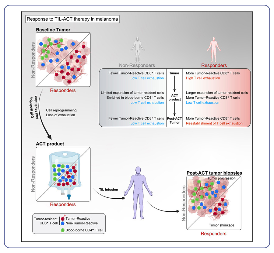
FDA has recently approved the first cancer tumor-infiltrating lymphocytes (TIL) therapy lifileucel (Amtagvi) for treatment of advanced melanoma. TIL (tumor-infiltrating lymphocyte) therapy, entails augmenting the population of immune cells within tumors, leveraging their potency to combat cancer6.

Genomics in skin cancer: In recent decades, advancements in skin cancer treatment, including targeted agents and immunotherapy, have significantly improved patient outcomes. Despite these strides, challenges persist, including limited efficacy, therapy resistance, and adverse effects. For example, while immunotherapy shows promise, it benefits only a subset of patients and can trigger adverse reactions. Combining therapies can enhance effectiveness but often leads to heightened side effects. Therefore, ongoing research aims to explore new targets, refine existing therapies, mitigate side effects, and comprehend resistance mechanisms. Genomic technologies, like next-generation sequencing (NGS), play a pivotal role in this pursuit. NGS panels have transformed skin cancer treatment by enabling precise molecular characterization. By analyzing individual patient tumor profiles, specific genetic alterations can be identified for targeted personalized therapies. This approach holds significant promise for enhancing treatment efficacy, minimizing side effects, and ultimately improving the prognosis for skin cancer patients.


Conclusions

The significant burden of skin cancer necessitates continued research efforts to improve prevention, early detection, and treatment strategies. This includes public health initiatives to promote sun protection and early detection measures, alongside advancements in personalized medicine through leveraging genetic insights. By tailoring treatment approaches based on individual patient characteristics and tumor profiles, we can strive for a future with more effective and less toxic therapies, ultimately reducing the morbidity and mortality associated with skin cancer.


MedGenome’s advanced solutions for skin cancer

MedGenome offers comprehensive tumor microenvironment solutions supported by a targeted sequencing approach, facilitating the identification of immune-oncology biomarkers such as microsatellite instability (MSI) and tumor mutational burden (TMB). Our optimized NGS assays enable the detection of low-prevalence pathogenic variants and epigenetic regulators relevant to various skin cancers. As pioneers in advanced genomic technologies and certified 10x service provider, we support your research journey from design to publication. Our expertise ensures the selection of optimal workflows, precise sample processing, and timely result delivery. We augment your research with custom visualizations, tailored analysis workflows, and seamless integration of external data, ensuring readiness for publication.

Feel free to contact our expert scientific team at research@medgenome.com for any questions or further information.

To know more about our advanced genomics solutions and services please click on the following links: Spatial Transcriptomics, Single cell sequencing, RNA Sequencing, Immune profiling, Whole genome and whole exome sequencing, and Epigenetic profiling


References


#Skin cancer, #Basal cell carcinoma, #Squamous cell carcinoma, #Melanoma, #UV radiation, #Melanocytes, #Mutations, #Treatment resistance, #Immunotherapy, #Targeted therapy, #Cellular therapy, #Clinical trials, #Genomics, #Skin cancer treatment


Unveiling the Complexity of Head and Neck Squamous Cell Carcinoma: From Genes to Microenvironment

Head and neck squamous cell carcinoma (HNSCC) ranks among the most prevalent cancers worldwide. In the United States, it is estimated that 58,450 new cases will be diagnosed in 2024, primarily affecting the oral cavity and pharynx1. Incidence rates among males are highest in non-Hispanic White and American Indian/Alaska Native individuals, with lower rates observed in Hispanic and Asian/Pacific Islander populations. Among females, incidence rates are elevated in non-Hispanic White and Asian/Pacific Islander individuals, while being lowest in Hispanic and Black populations.

By MedGenome Scientific Affairs

Head and neck squamous cell carcinoma (HNSCC) ranks among the most prevalent cancers worldwide. In the United States, it is estimated that 58,450 new cases will be diagnosed in 2024, primarily affecting the oral cavity and pharynx1. Incidence rates among males are highest in non-Hispanic White and American Indian/Alaska Native individuals, with lower rates observed in Hispanic and Asian/Pacific Islander populations. Among females, incidence rates are elevated in non-Hispanic White and Asian/Pacific Islander individuals, while being lowest in Hispanic and Black populations. Major risk factors for HNSCC include tobacco and alcohol use, as well as Human Papilloma Virus (HPV) infection. While tobacco-related HNSCC rates have declined over time, rising incidence rates, particularly among younger individuals, are attributed to HPV-related disease2.

Molecular pathogenesis of HNSCC

HNSCC, like other solid tumors, develops through genetic and epigenetic alterations, leading to various cancer phenotypes. Whole exome sequencing analyses of HNSCC specimens have identified mutations targeting key oncogenes and tumor suppressor pathways, such as p53, Rb/INK4/ARF, and Notch, which regulate cellular processes like proliferation, differentiation, and metastasis. Studies, including The Cancer Genome Atlas (TCGA) project, have categorized HNSCC based on genetic and expression patterns, revealing distinct subtypes with unique molecular features and clinical characteristics. Notably, mutations in genes like TP53 and CDKN2A are prevalent, while HPV+ tumors exhibit different mutation profiles, with frequent amplifications in PIK3CA and SOX2 genes. Furthermore, the Notch signaling pathway and PIK3CA alterations have been implicated in HNSCC progression and immune evasion3.

Tumor microenvironment of HNSCC

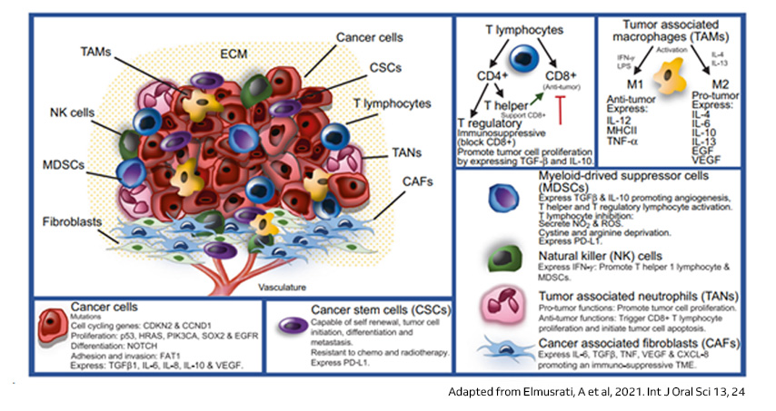
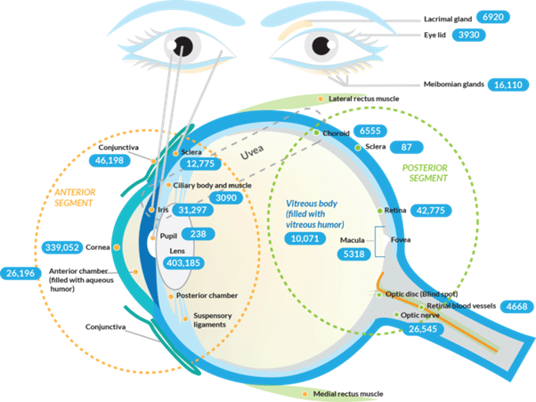
The tumor microenvironment (TME) in HNSCC and cancer generally comprises a diverse mix of cancer cells and nonmalignant components, including immune cells like T lymphocytes, tumor associated macrophages (TAMs), myeloid-derived suppressor cells (MDSCs), NK cells, tumor associated neutrophils (TANs), and Cancer associated fibroblasts (CAFs), along with fibroblasts, mesenchymal cells, and vascular endothelial cells. These nonmalignant cells play dual roles in tumor growth and dissemination. Understanding the immune landscape within the TME is crucial for assessing cancer progression and the efficacy of immunotherapy. Research on the TME in HNSCC highlights its impact on cancer behavior through complex cellular interactions mediated by growth factors and cytokines. Inflammatory processes within the TME worsen malignancy, with various immune cells exhibiting diverse functions affecting prognosis. Additionally, cytokines like TGFβ and IL-6 contribute to immunosuppression, influencing therapeutic responses, while hypoxia further promotes immune evasion and tumor progression3,4.

Illustration of the tumor microenvironment in HNSCC
Figure 1: Illustration of the tumor microenvironment in HNSCC

Single cell sequencing and its applications in HNSCC

Intra-tumoral heterogeneity (ITH) and plasticity present significant hurdles in translating cancer research into effective therapies due to the varied cellular compositions and dynamic functional states within tumors. Recent advances in single-cell sequencing have substantially improved the resolution of studies exploring ITH, the TME, and intra-tumoral cell-cell communication. This technology allows for the analysis of individual cells within the TME, revealing previously unseen diversity and dynamics. By identifying rare cell populations, characterizing cellular interactions, and discovering novel biomarkers and therapeutic targets, single-cell sequencing has the potential to revolutionize our understanding of HNSCC biology and pave the way for personalized diagnostic and therapeutic strategies. Some of the key highlights of single cell sequencing studies in HNSCC are provided below.

Insights into tumor composition and behavior

In one of the initial single cell sequencing studies on oral squamous cell carcinoma (OSCC), distinct non-malignant cell clusters were identified, including T-cells, macrophages, fibroblasts, and others, with T-cell subsets exhibiting variable sizes across patients. Conversely, malignant cells displayed patient-specific clustering, with only a few shared signatures among tumors, notably featuring a partial Epithelial-mesenchymal transition (EMT) program associated with advanced disease characteristics. Subsequent investigations involving a wider range of HNSCC subsites and HPV statuses corroborated these findings. These studies identified additional subclusters of fibroblasts and revealed patient-specific clustering patterns of malignant cells, with a correlation observed between these patterns and HPV status. Further exploration into cancer stem cells through scRNAseq highlighted metabolic program variations and extensive ITH across multiple tumor types, suggesting that transcriptional ITH reflects tissue heterogeneity5,6.

Decoding the complexity of TME

The immune system serves as a critical defense mechanism against malignant cells, leading to the development of immunotherapy as a prominent treatment strategy in cancer. However, despite its approval for HNSCC, immune checkpoint inhibitors only benefit a minority of patients. To understand the intricacies of the HNSCC TME, several studies have utilized single-cell RNA sequencing (scRNAseq) on sorted cell populations, including CD45+ hematopoietic cells or CD3+ T-cells. These studies often incorporate adjacent normal tissue, peripheral blood leukocytes, and non-tumorous tonsils for comparison. Analyses primarily focused on T-cells due to their pivotal role in antitumor immunity and immunotherapy. These studies revealed distinct T-cell subsets, such as CD8+ cells, which were found in higher proportions within tumor tissue compared to adjacent normal tissue. Additionally, investigations into CD4+ T-cells highlighted an increased presence of regulatory T-cells (Tregs) within tumors, indicative of an immunosuppressive tumor microenvironment. Furthermore, studies identified potential routes of cell-cell communication, particularly emphasizing the interaction between macrophages and T-cells mediated by PD-L1. Mouse models and scRNAseq data also showed how immune responses within tumors can vary, with T-cells multiplying in specific ways. This provided insights into tumor antigen recognition and how immune responses differ between patients. Explorations into the humoral arm of anti-tumor immunity and the contribution of natural killer (NK) cells underscored their potential therapeutic implications in HNSCC5,6.


Conclusions

HNSCC presents a complex interplay between genetics, the tumor microenvironment, and the immune system. Understanding these factors is crucial for developing effective therapeutic strategies. Single-cell sequencing has emerged as a powerful tool, shedding light on the intricate cellular diversity within HNSCC tumors and the dynamic interactions within the TME. These insights not only hold promise for personalized medicine in HNSCC but also contribute significantly to our overall understanding of cancer biology, paving the way for advancements in cancer research across different tumor types.


MedGenome solutions

As a 10x certified service provider and an early pioneer in single-cell genomic sequencing, MedGenome offers comprehensive support throughout your research journey, from experimental design to publication. Our expertise spans selecting the most suitable single-cell workflow, processing diverse sample types with efficiency and accuracy, and delivering timely results. With custom visualizations, tailored analysis workflows, and seamless integration of external data, we ensure your research is publication-ready. Our proprietary algorithm, OncoPeptTUMETM utilizes RNA-seq data to create high-resolution maps of the tumor microenvironment based on specific cell type gene signatures.

For any queries or additional details, please reach out to our expert scientific team at research@medgenome.com.

To know more about our unique cancer genomics solutions and services please click on the following links: Whole genome and whole exome sequencing, RNA Sequencing, Single cell sequencing, Immune profiling and Epigenetic profiling


References

    • Siegel RL, Giaquinto AN, Jemal A. Cancer statistics 2024 (2023). CA Cancer J Clin. 2024. 74(1):12-49.
    • Barsouk A, Aluru JS, Rawla P, Saginala K, Barsouk A. Epidemiology, Risk Factors, and Prevention of Head and Neck Squamous Cell Carcinoma (2023). Med Sci (Basel). 13;11(2):42
    • Elmusrati, A., Wang, J. & Wang, CY. Tumor microenvironment and immune evasion in head and neck squamous cell carcinoma (2021). Int J Oral Sci 13, 24.
    • Ruffin AT, Li H, Vujanovic L, Zandberg DP, Ferris RL, Bruno TC. Improving head and neck cancer therapies by immunomodulation of the tumour microenvironment. Nat Rev Cancer. 23(3):173-188.
    • Qi Z, Barrett T, Parikh AS, Tirosh I, Puram SV. Single-cell sequencing and its applications in head and neck cancer (2019). Oral Oncol. 99:104441.
    • Heller G, Fuereder T, Grandits AM, Wieser R. New perspectives on biology, disease progression, and therapy response of head and neck cancer gained from single cell RNA sequencing and spatial transcriptomics (2023) Oncol Res. 32(1):1-17.


#Head and Neck cancer, #Oral squamous cell carcinoma, #Tumor microenvironment, #Single cell sequencing, #Immunotherapy, #Immune checkpoint inhibitors, #Intra-tumoral heterogeneity, #Epithelial-mesenchymal transition, #Human papilloma virus